
Teacher Portal
Investigation 1: Concepts
Mitosis and Human Chromosomes
Navigate:
Once the slide presentation is launched
- use your left and right arrows to advance or go back in the slide presentation, and
- hover your mouse over the left edge of the presentation to get a view of the thumbnails for all the slides so that you can quickly move anywhere in the presentation.
- Click HERE to launch the slide presentation for the CELL.
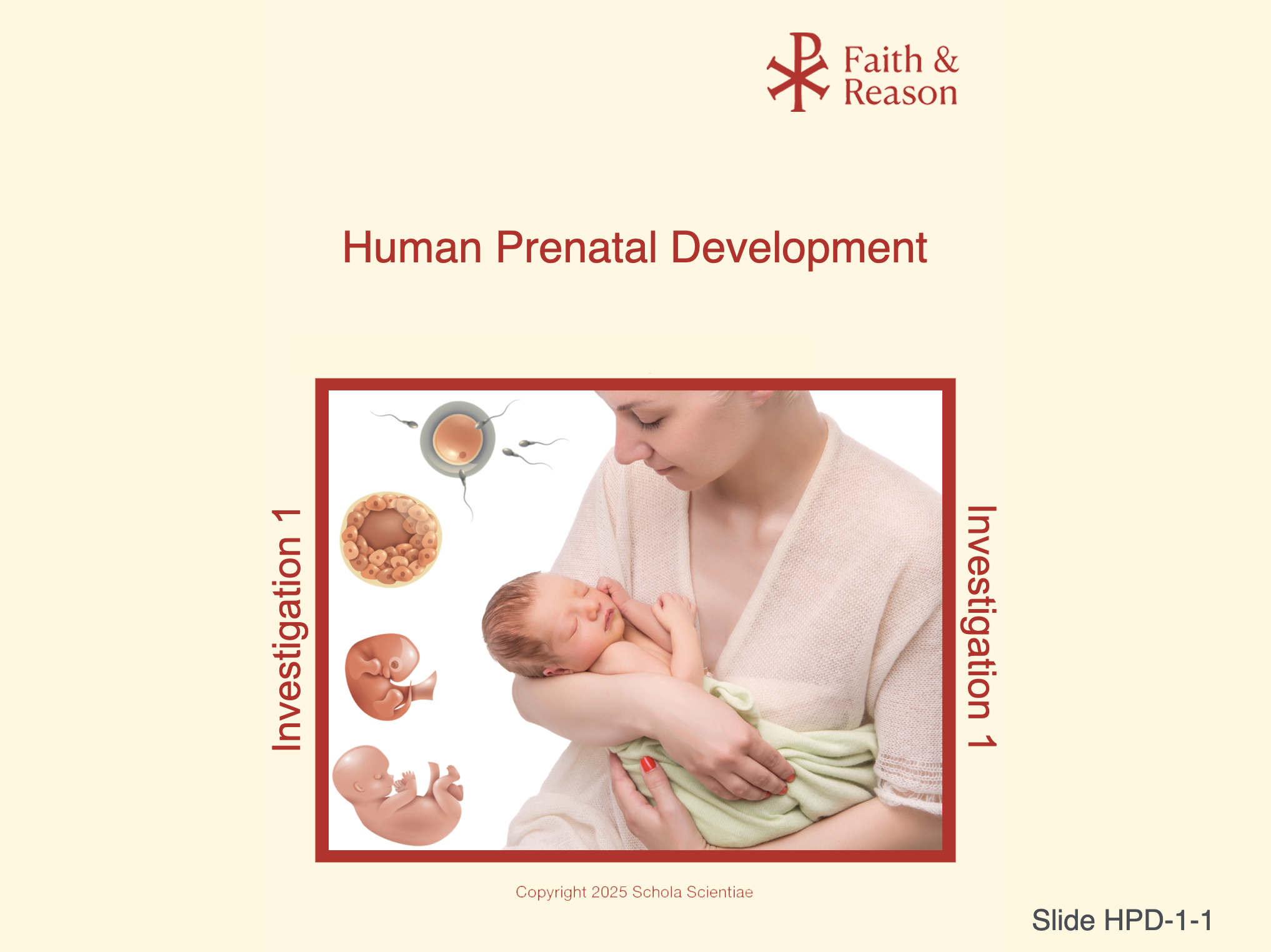
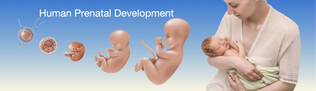
SLIDE HPD-1-1 Human Chromosomes and Mitosis
Purpose of This Slide
This opening slide introduces the central scientific focus of Investigation 1: how chromosomes carry genetic information and how mitosis allows that information to be faithfully passed on as a human being grows and develops.
Use this slide to orient students to what kind of investigation this is:
It is evidence-based
It builds from observable cellular structures
It explains growth from a single cell to a multicellular human organism
This slide is not meant to teach details yet. Its purpose is to establish scope, seriousness, and coherence.
Key Points to Emphasize
Human development depends on accurate genetic information
Chromosomes are the structures that carry this information
Mitosis is the process that allows cells to divide while keeping genetic instructions the same
This investigation will connect cell biology to human growth and development
Discussion Questions (with Suggested Answers)
Question 1: What are chromosomes, and why are they important?
Suggested Answer: Chromosomes are organized structures made of DNA that carry genetic instructions needed for growth, development, and function.
Question 2: What is mitosis, in general terms?
Suggested Answer: Mitosis is the process by which a cell divides to produce two genetically identical cells.
Question 3: Why is it important to understand these processes when studying human development?
Suggested Answer: Because human growth depends on cells dividing accurately and consistently from the earliest stages of life.
Pedagogical Note
Resist the urge to preview too many details. Let students know that clarity will come through visuals, discussion, and lab work. This slide should create confidence, not cognitive overload.
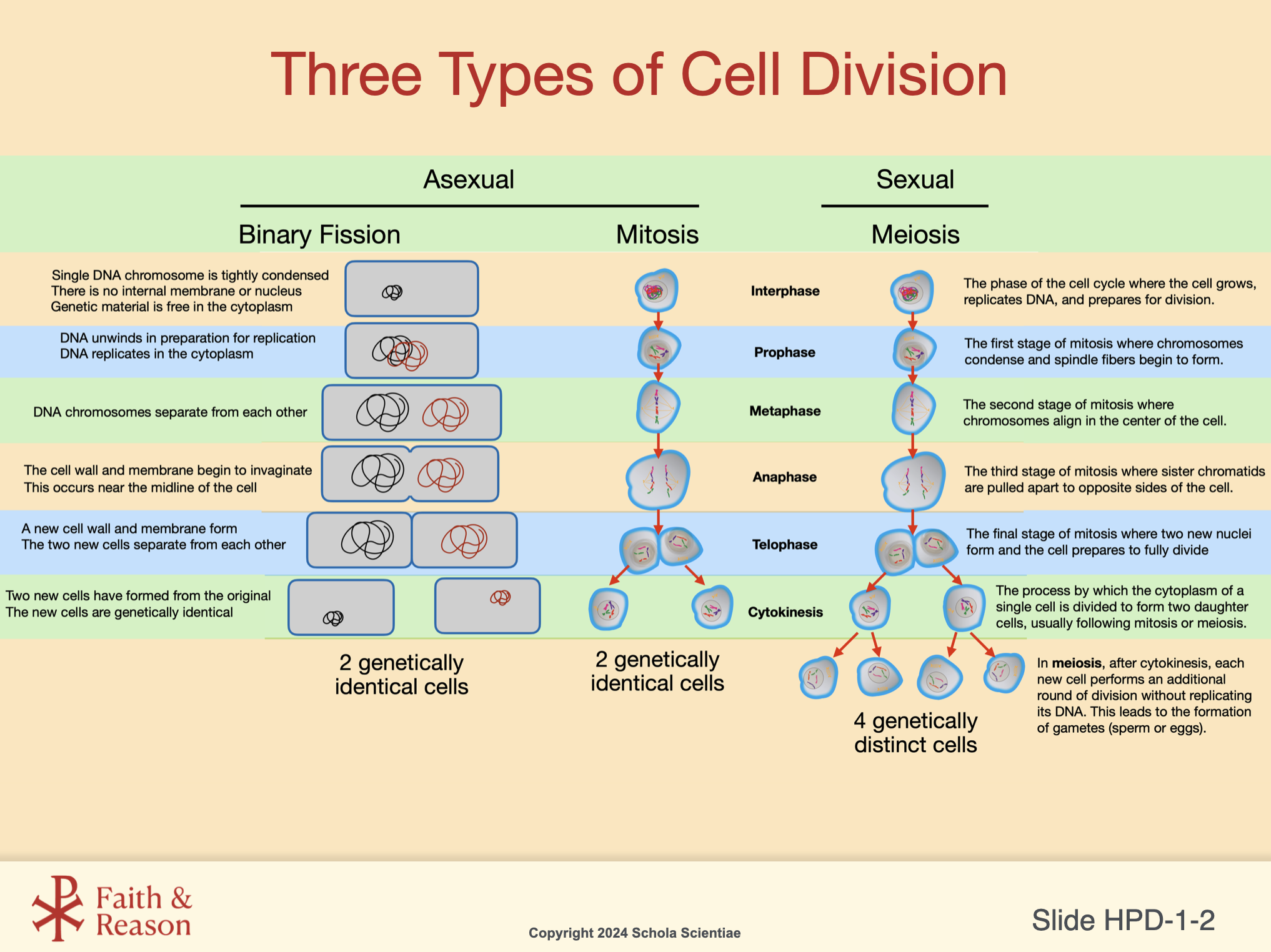
SLIDE HPD-1-2 Three Types of Cell Division
Purpose of This Slide
This slide provides a conceptual overview of three different types of cell division: binary fission, mitosis, and meiosis. Its purpose is not to teach details, but to help students see that cell division occurs in different biological contexts and serves different purposes.
Use this slide to orient students to the idea that:
Not all cell division is the same
Different organisms divide cells in different ways
Human growth depends specifically on mitosis, which will be the focus of this investigation
This slide establishes context and comparison, not mastery.
Key Points to Emphasize
Binary fission is a form of asexual reproduction used by single-celled organisms
Mitosis produces two genetically identical cells used for growth and repair
Meiosis produces genetically distinct cells used for sexual reproduction
All three processes involve copying and distributing genetic information
Only mitosis explains how a human body grows from one cell to many
Discussion Questions (with Suggested Answers)
Question 1: Why might different organisms use different types of cell division?
Suggested Answer: Because different organisms have different structures and reproductive needs.
Question 2: Which type of cell division is most important for human growth and development?
Suggested Answer: Mitosis, because it produces genetically identical cells for growth and repair.
Question 3: Why do you think it is useful to compare these processes before studying mitosis in detail?
Suggested Answer: Comparing them helps students understand what makes mitosis distinct and why it is biologically important.
Pedagogical Note
Avoid detailed stage-by-stage explanations here. Students will return to mitosis repeatedly. This slide should reduce confusion, not create it.
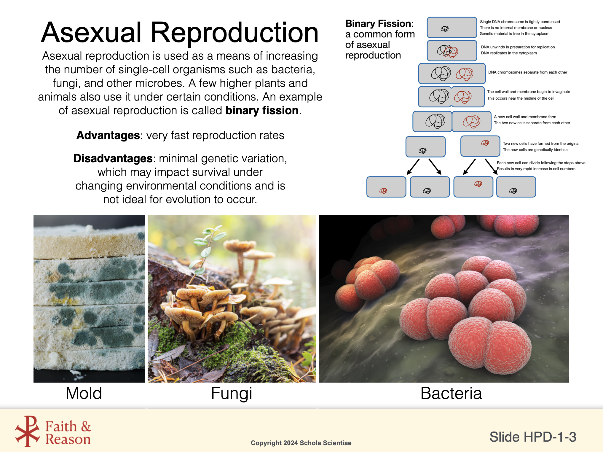
SLIDE HPD-1-3 Asexual Reproduction (Binary Fission)
Purpose of This Slide
This slide introduces binary fission as a form of asexual reproduction used by single-celled organisms such as bacteria. Its purpose is to show how cell division can function as reproduction, not just growth.
This contrast is essential:
Binary fission = reproduction of an entire organism
Mitosis = growth and maintenance within a multicellular organism
Students must understand this distinction to avoid confusion later.
Key Points to Emphasize
Binary fission is used by single-celled organisms
The process produces genetically identical cells
It allows for rapid reproduction
Lack of genetic variation can be a disadvantage in changing environments
Humans do not reproduce by binary fission
Discussion Questions (with Suggested Answers)
Question 1: What is binary fission?
Suggested Answer: Binary fission is a form of asexual reproduction in which one cell divides into two genetically identical cells.
Question 2: What types of organisms use binary fission?
Suggested Answer: Single-celled organisms such as bacteria.
Question 3: Why is binary fission not used for human reproduction or growth?
Suggested Answer: Because humans are multicellular organisms that require mitosis for growth and meiosis for reproduction.
Pedagogical Note
Reinforce the idea that similar processes can have very different biological meanings, depending on the organism. This prepares students for careful thinking later.
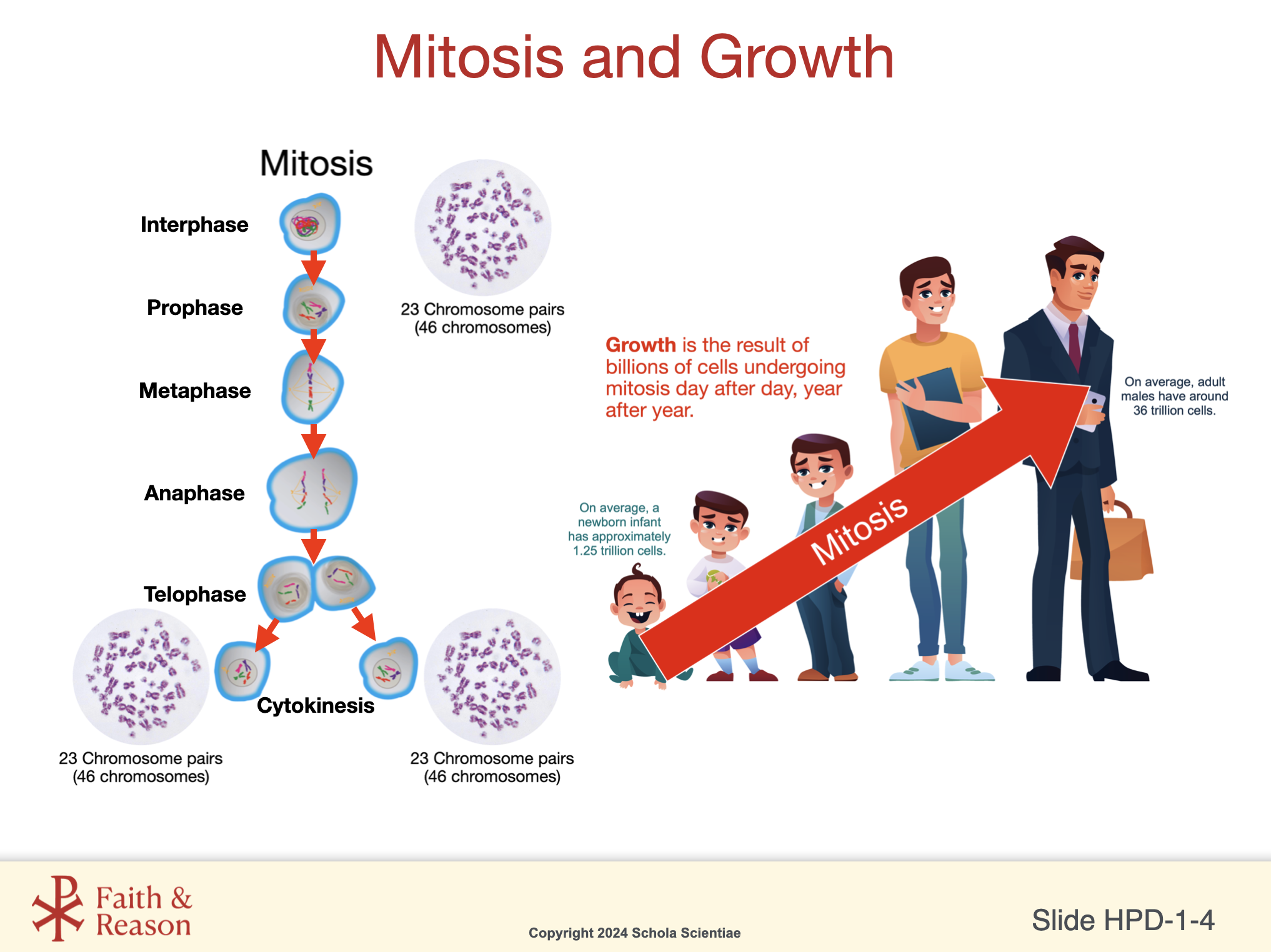
SLIDE HPD-1-4 Mitosis: Cell Division for Growth and Repair
Purpose of This Slide
This slide introduces mitosis as the type of cell division used by multicellular organisms, including humans, for growth, development, and repair. Its purpose is to clearly distinguish mitosis from binary fission and to establish mitosis as the central mechanism of this investigation.
Use this slide to emphasize that:
Mitosis does not create a new organism
Mitosis increases the number of cells within an existing organism
Genetic information must be copied and preserved accurately for normal development
This slide marks the transition from comparison to focus.
Key Points to Emphasize
Mitosis is used by multicellular organisms
It produces two genetically identical cells
These cells are used for growth, repair, and replacement
Accurate copying of genetic information is essential
Human development depends on mitosis beginning at the earliest stages of life
Discussion Questions (with Suggested Answers)
Question 1: What is mitosis, in general terms?
Suggested Answer: Mitosis is a process in which one cell divides to form two genetically identical cells.
Question 2: How is mitosis different from binary fission?
Suggested Answer: Binary fission is a form of reproduction in single-celled organisms, while mitosis supports growth and repair in multicellular organisms.
Question 3: Why is it important that cells produced by mitosis are genetically identical?
Suggested Answer: Because identical genetic information allows tissues and organs to grow and function normally.
Pedagogical Note
Avoid detailed phase names at this point. Students will encounter the stages of mitosis later. Keep the focus on purpose and outcome, not sequence.
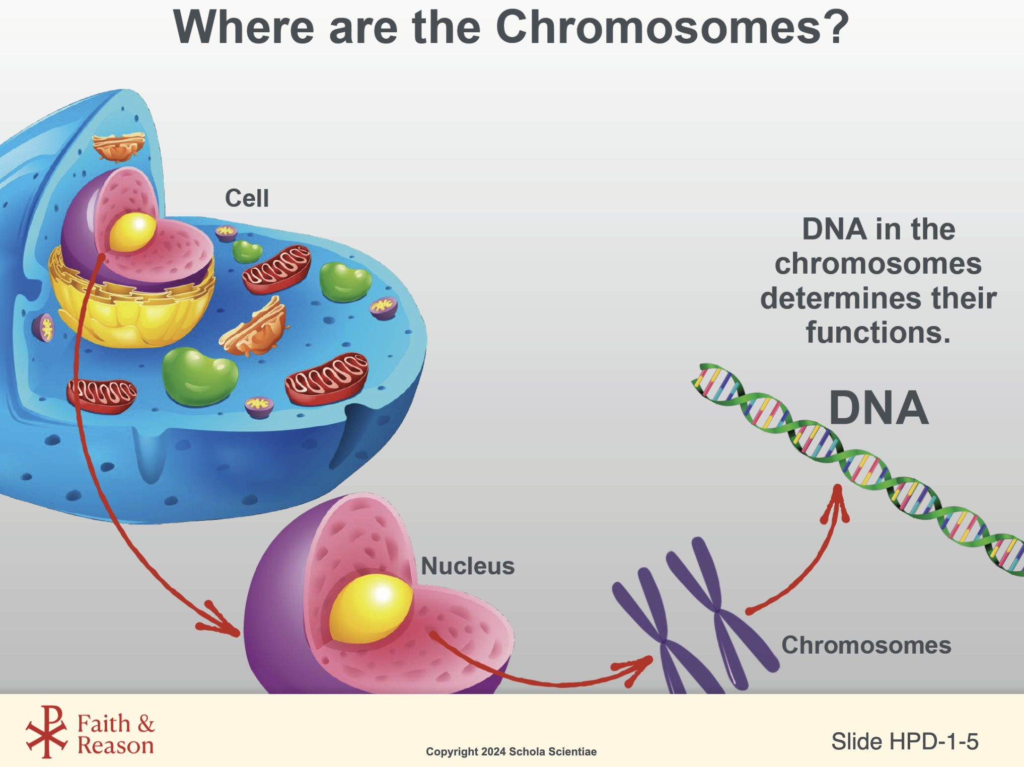
SLIDE HPD-1-5 The Nucleus and Chromosomes
Purpose of This Slide
This slide establishes where genetic information is physically located in the cell and how it is protected and organized. Emphasize that chromosomes are real, observable structures, not abstract ideas. This will later support discussions of inheritance, development, and continuity from conception.
Students often confuse:
“DNA” with “chromosomes”
“genes” with “traits”
“instructions” with outcomes
Use this slide to clarify that DNA is organized into chromosomes, which reside in the nucleus, and that accurate copying of this information is essential for normal development.
Key Points to Emphasize
The nucleus protects genetic information.
Chromosomes are organized packages of DNA.
DNA must be copied before cell division so each new cell receives the same instructions.
This organization is foundational for growth, repair, and development.
Discussion Questions (with Suggested Answers)
Question 1: Where are chromosomes located inside a human cell?
Suggested Answer: Chromosomes are located inside the nucleus.
Question 2: What is the role of DNA within chromosomes?
Suggested Answer: DNA carries the genetic instructions that guide how cells grow, function, and develop.
Question 3: Why must DNA be copied before a cell divides?
Suggested Answer: So that each new cell receives a complete and identical set of genetic instructions, allowing normal growth and function.
Pedagogical Note
Avoid implying that DNA determines everything about a person. Reinforce that DNA provides instructions, while development unfolds through complex biological processes that will be explored throughout the unit.
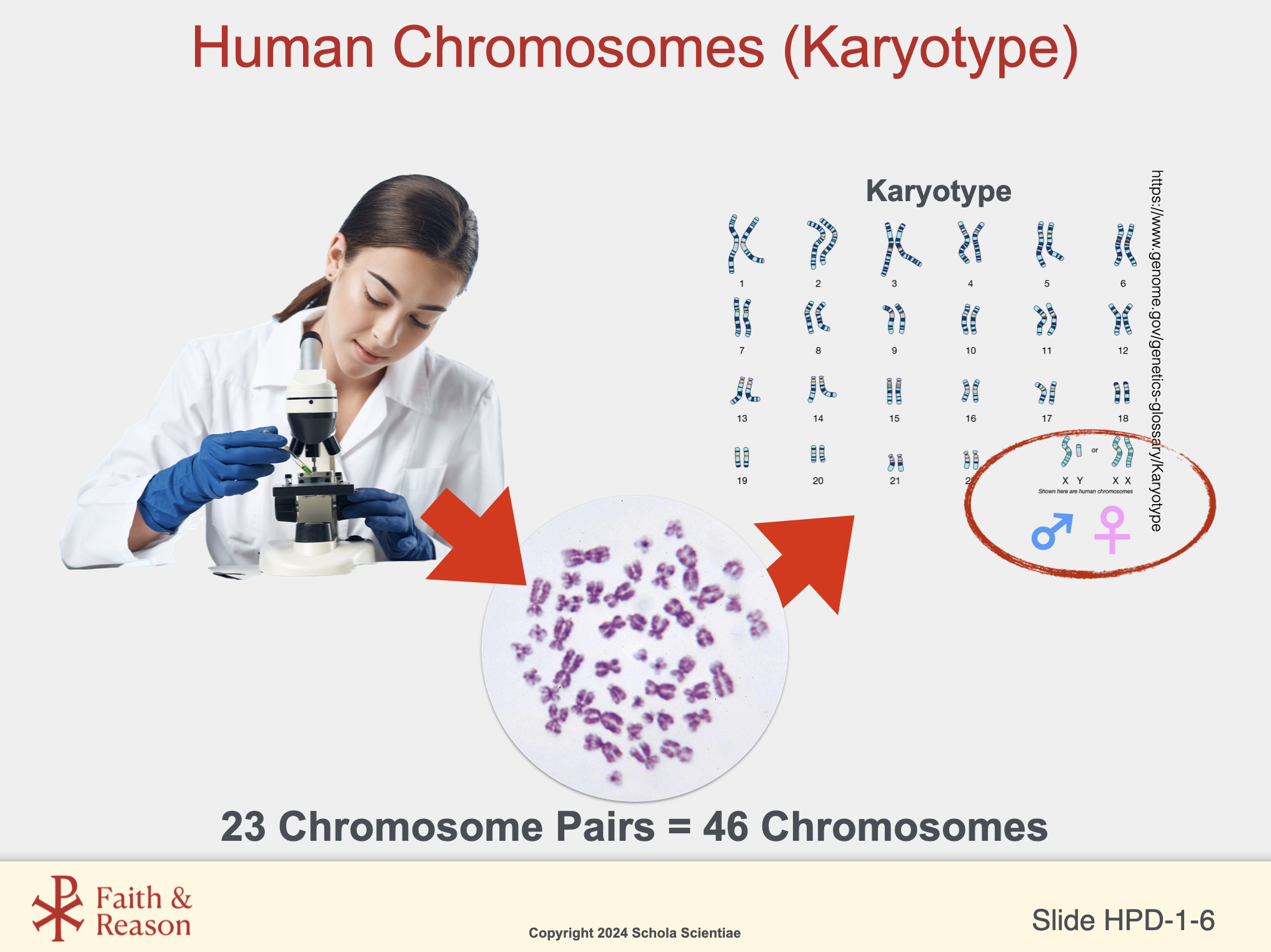
SLIDE HPD-1-6 Human Chromosomes
Purpose of This Slide
This slide introduces karyotypes as a scientific tool used to organize and study human chromosomes. Emphasize that chromosomes are physical structures that can be observed, photographed, counted, and compared. This reinforces that genetics is grounded in direct observation, not abstraction.
This slide also establishes the biological basis of chromosome number and pairing, which will later support discussions of inheritance, variation, and sex determination in Investigation 2.
Key Points to Emphasize
Humans have 46 chromosomes, arranged as 23 pairs.
One chromosome in each pair comes from the mother, the other from the father.
A karyotype is a visual arrangement of chromosomes used by scientists.
Karyotypes allow scientists to:
Confirm chromosome number
Identify sex chromosomes (XX or XY)
Detect certain genetic conditions
Discussion Questions (with Suggested Answers)
Question 1: How many chromosomes are found in most human cells?
Suggested Answer: Humans have 46 chromosomes, arranged in 23 pairs.
Question 2: What is a karyotype, and why do scientists use it?
Suggested Answer: A karyotype is an organized chart of chromosomes that scientists use to study chromosome number, structure, and genetic conditions.
Question 3: What determines whether a person is biologically male or female at the chromosome level?
Suggested Answer: The sex chromosomes. XX typically indicates female, and XY typically indicates male. The father’s sperm contributes either an X or a Y chromosome.
Pedagogical Note
Avoid extending this slide into sociological or ethical discussions. Keep the focus on biological description, not interpretation. This scientific clarity is essential before any later reflection on meaning, dignity, or moral questions.
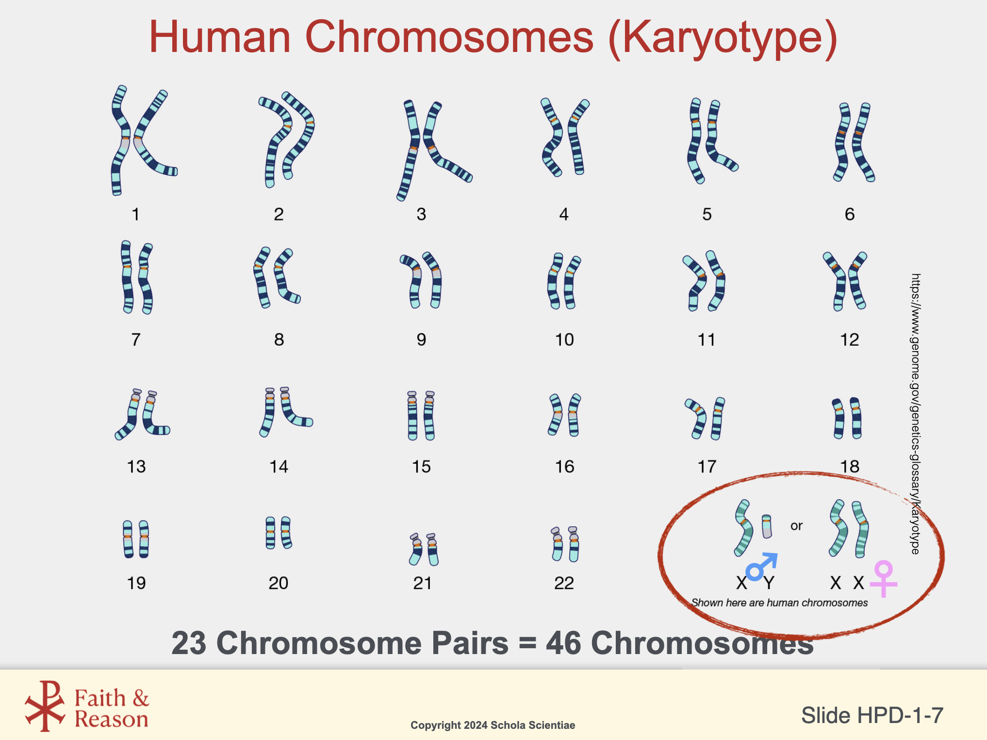
SLIDE HPD-1-7
Purpose of This Slide
This slide presents a complete human karyotype, showing all 23 pairs of chromosomes arranged and numbered. Its purpose is to reinforce that chromosome number, pairing, and structure are observable biological facts, not theoretical constructs.
This slide also makes explicit that one chromosome pair—the sex chromosomes—differs between males and females, establishing the biological basis of sex determination at the chromosomal level.
Use this slide to consolidate earlier ideas:
Chromosomes are physical structures
They are inherited in pairs
They can be studied, compared, and identified
Key Points to Emphasize
Humans have 46 chromosomes, arranged as 23 pairs
Chromosome pairs 1–22 are called autosomes
The 23rd pair consists of the sex chromosomes
Typical sex chromosome combinations are:
XX (female)
XY (male)
This chromosomal arrangement is established at conception
Discussion Questions (with Suggested Answers)
Question 1: What does a karyotype show scientists?
Suggested Answer: A karyotype shows the number, size, and pairing of chromosomes in a cell.
Question 2: How many chromosome pairs do humans have, and how many total chromosomes?
Suggested Answer: Humans have 23 pairs of chromosomes, for a total of 46.
Question 3: What is the role of the sex chromosomes in human development?
Suggested Answer: The sex chromosomes help determine biological sex at the chromosomal level, with XX typically female and XY typically male.
Pedagogical Note
Maintain focus on biological description, not interpretation. Avoid extending this slide into sociological, psychological, or ethical discussions. Scientific clarity here is essential groundwork for later reflection handled elsewhere in the curriculum.
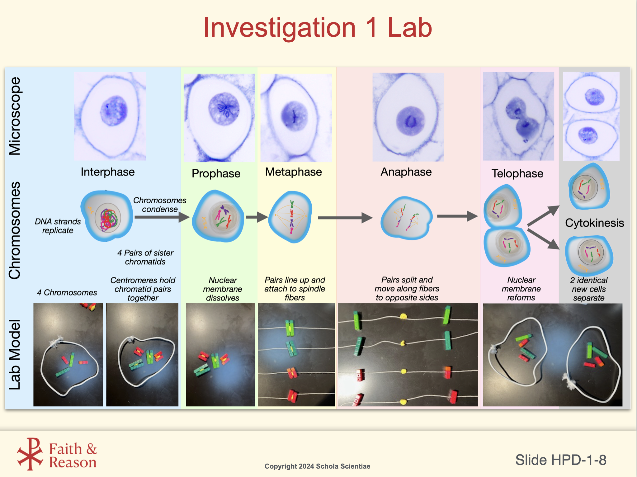
SLIDE HPD-1-8 Investigation 1 Lab: Mitosis
Purpose of This Slide
This slide integrates three representations of mitosis into a single, coherent sequence:
Microscope images (real cells)
Chromosome diagrams (simplified conceptual models)
Hands-on lab models (student-constructed representations)
The purpose is to help students recognize that mitosis is a real, observable biological process that can be understood consistently across multiple levels of representation.
This slide serves as the conceptual bridge between:
What scientists observe
What diagrams explain
What students physically model in the lab
Key Points to Emphasize
Mitosis is a process, not a single event
DNA is replicated before mitosis begins (interphase)
Chromosomes condense, align, separate, and are re-enclosed in nuclei
Sister chromatids separate, not chromosome pairs
Cytokinesis completes cell division, producing two genetically identical daughter cells
Emphasize correct sequencing:
Interphase → Prophase → Metaphase → Anaphase → Telophase → Cytokinesis
Discussion Questions (with Suggested Answers)
Question 1: Why is interphase included even though it is not technically part of mitosis?
Suggested Answer: Because DNA replication occurs during interphase, which is essential for mitosis to produce identical daughter cells.
Question 2: What is separating during anaphase?
Suggested Answer: Sister chromatids separate and move to opposite sides of the cell.
Question 3: How does cytokinesis differ from mitosis?
Suggested Answer: Mitosis divides the nucleus, while cytokinesis divides the cytoplasm to form two separate cells.
Pedagogical Note
Students often confuse chromosome number with chromatid number. Use the lab model images to slow this down and reinforce that chromosome number remains constant through mitosis.
Avoid premature comparisons to meiosis on this slide. Precision here prevents major misconceptions later.
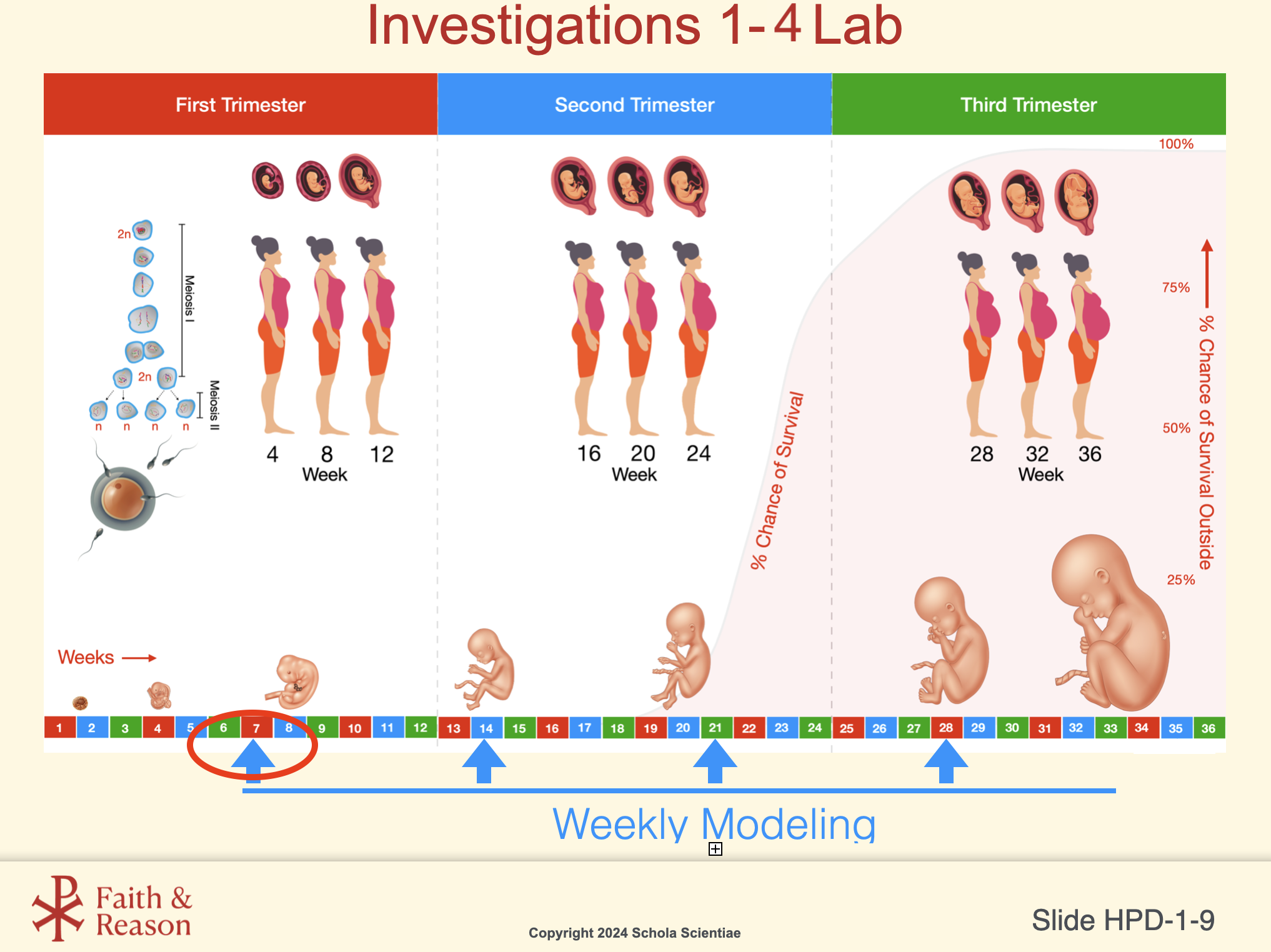
SLIDE HPD-1-9 Investigations 1–4 Lab: Weekly Modeling of Prenatal Development
Purpose of This Slide
This slide establishes the longitudinal framework for the entire Human Prenatal Development CELL. It visually connects:
Time (weeks of pregnancy)
Growth (length and mass)
Developmental stages (trimesters)
Biological viability (survival outside the womb)
Students will return to this slide repeatedly across Investigations 1–4, using it as a reference for data-driven modeling and discussion.
Key Instructional Roles of This Slide
Anchors all lab modeling activities to real prenatal data
Reinforces that prenatal development is continuous and measurable
Helps students connect microscopic processes (cell division) to macroscopic outcomes (growth and survival)
Provides a shared visual reference across multiple investigations
Top Section: The Three Trimesters
First Trimester (Weeks 1–12): Major organs begin forming
Second Trimester (Weeks 13–26): Rapid growth and increasing movement
Third Trimester (Weeks 27–40): Weight gain and physiological preparation for birth
Emphasize that the fetus is the same developing individual throughout all three trimesters.
Bottom Section: Weekly Modeling Activity
Students model fetal length and mass at weeks 7, 14, 21, and 28
The color-coded timeline represents week-by-week development
Blue arrows indicate specific weeks used for hands-on modeling
Stress accuracy and scale: models are not symbolic — they represent real biological measurements.
Right Side: Survival Outside the Womb
The red curve shows percent chance of survival if birth occurs early
Survival remains very low before approximately week 22
Survival increases rapidly as organs — especially the lungs — mature
This data must be treated carefully and scientifically, without speculation or moral commentary at this stage.
Discussion Questions (with Suggested Answers)
Question: What will students be modeling throughout the CELL?
Suggested Answer: Fetal growth using real length and mass data at weeks 7, 14, 21, and 28.
Question: Why does survival outside the womb increase later in pregnancy?
Suggested Answer: Because organ systems, especially the lungs, become more fully developed.
Question: How does this slide connect earlier topics like mitosis and meiosis to later development?
Suggested Answer: Cell division enables growth, which leads to measurable changes in size, structure, and viability.
Pedagogical Note
This slide is intentionally revisited. Repetition here is a feature, not a flaw — it builds conceptual continuity, reduces cognitive load, and reinforces scientific reasoning through time-based data.
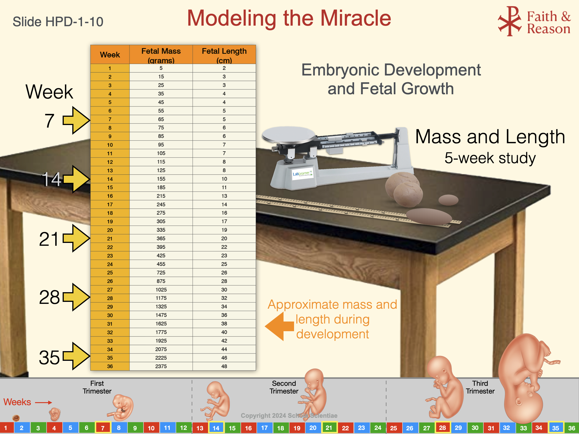
SLIDE HPD-1-10 Modeling the Miracle
Purpose of This Slide
This slide introduces the central hands-on lab experience of the Human Prenatal Development CELL. Students will model fetal growth by tracking mass and length over time, using real biological data.
The lab is intentionally quantitative, physical, and cumulative. It transforms abstract growth curves into measurable, tangible experience, reinforcing that prenatal development is:
Continuous
Predictable
Data-driven
Scientifically observable
Left Section: Fetal Growth Data Table
The table provides realistic fetal mass and length values from weeks 1–36
Highlighted weeks (7, 14, 21, 28, and 35) indicate when students will construct models
Students use this data to scale their clay models accurately
Emphasize that these numbers are not estimates invented for class, but values derived from medical and developmental research.
Right Section: Lab Setup
Students use rulers and balances to measure length and mass
The image of the triple-beam balance represents the measurement mindset of the lab
Precision matters: students must revise models if measurements do not match data
This is not an art project. It is a biological modeling exercise grounded in measurement and verification.
Bottom Section: Weekly Growth Timeline
The timeline situates fetal growth within trimesters
Students will refer to this visual repeatedly across Investigations 1–3
Models are compared against the timeline to identify patterns of growth acceleration
Discussion Questions (with Suggested Answers)
Question: What are students modeling in this lab?
Suggested Answer: Fetal growth in terms of length and mass at specific weeks of development.
Question: Why is it important to measure both length and mass?
Suggested Answer: Because growth involves changes in size and weight, reflecting different aspects of development.
Question: How does this lab connect earlier topics like mitosis to later development?
Suggested Answer: Cell division enables growth, which results in measurable increases in mass and length over time.
Pedagogical Note
This lab deliberately slows students down. By requiring repeated modeling and comparison to real data, students confront the scale, rate, and continuity of prenatal development in a way that numbers or diagrams alone cannot achieve.
The lab prepares students intellectually for later reflection — without requiring that reflection yet.