
Teacher Portal
Investigation 1: PreLab
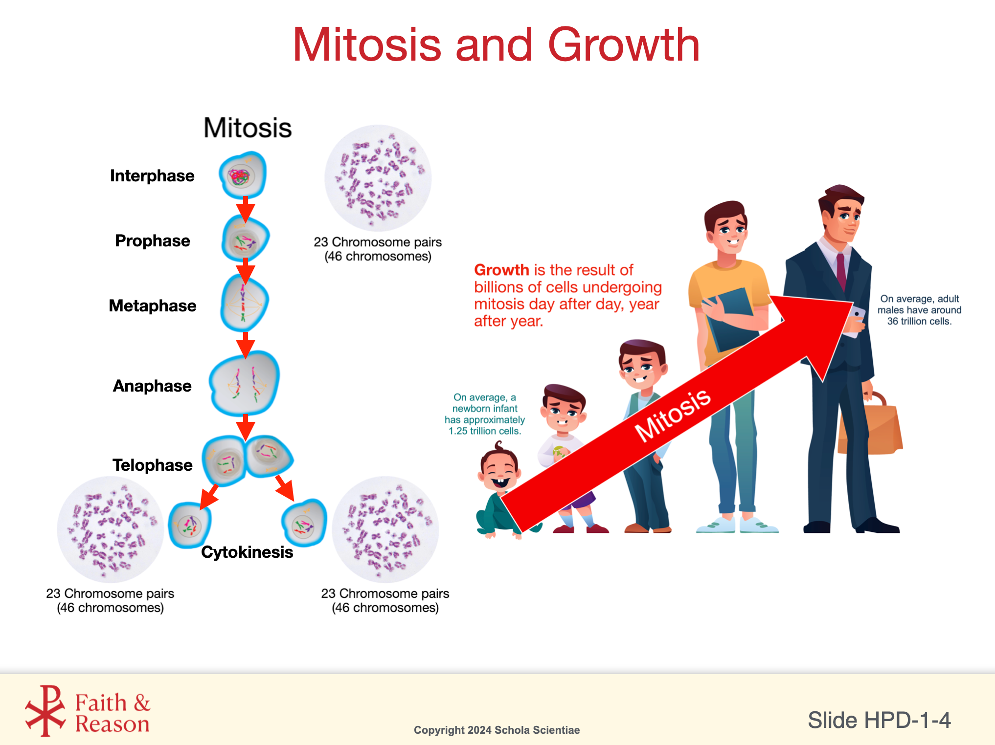
Mitosis and Human Chromosomes
Purpose of the PreLab
During this PreLab, students prepare for the upcoming laboratory investigation by reviewing and organizing key concepts related to mitosis and human chromosomes. This preparation helps students approach the lab with clear expectations and a stronger understanding of how controlled cell division supports prenatal development.
Focus Questions:
1. How does human prenatal development depend on orderly, precisely controlled biological processes?
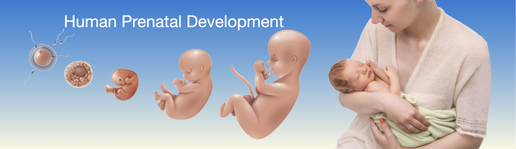
Human prenatal development depends on a sequence of biological processes that occur in a precise, regulated, and highly coordinated manner. From the moment of fertilization, development proceeds through predictable stages that rely on accurate cell division, controlled gene expression, and regulated cell differentiation. Cells do not divide randomly; they follow internal instructions that determine when to divide, how often to divide, and what type of cell to become.
This orderliness allows complex structures—such as tissues, organs, and body systems—to form in the correct locations and proportions. Even small deviations from these regulated processes can disrupt development, which highlights how tightly controlled prenatal growth must be. The remarkable reliability of human development across billions of individuals underscores that these processes are governed by biological laws rather than chance.
Discussion angles students may raise:
“Everything happens in steps.”
“Cells have instructions.”
“It’s not random.”
“Things grow in the right order.”
Teacher move:
Affirm these ideas and emphasize control, regulation, and coordination.
 Clarity Tool #2: Data vs Interpretation
This is a good moment to help students distinguish what they observe from the conclusions they begin to draw.
2. How can a single fertilized cell give rise to the trillions of cells in a human body?
A single fertilized cell gives rise to trillions of cells through repeated cycles of mitosis, in which one cell divides into two genetically identical cells. Each of these cells can then divide again, leading to exponential growth in cell number. Early in development, these divisions happen rapidly, allowing the embryo to grow from a microscopic single cell into a multicellular organism.
Importantly, this increase in cell number does not occur all at once but through countless rounds of controlled division. As development progresses, cells also begin to specialize, forming different tissues and organs, even though they all originated from the same original cell. This process shows how growth depends on both cell multiplication and cell organization.
Discussion angles students may raise:
“Cells keep dividing.”
“It doubles each time.”
“Cells start the same but become different.”
“Growth happens gradually.”
Teacher move:
Reinforce the idea of exponential increase + regulation, not just “more cells.”
3. How does mitosis allow cells in a developing embryo to remain genetically identical while increasing in number?
Mitosis ensures that each new cell receives an exact copy of the original cell’s genetic material. Before division, the cell duplicates its chromosomes so that two identical sets are present. During mitosis, these duplicated chromosomes are carefully separated and distributed evenly into two daughter cells. As a result, each new cell contains the same genetic information as the original cell, allowing the embryo to grow while maintaining genetic consistency across all cells.
Student Discussion Angles
Focus on chromosome duplication and separation
Connect chromosome behavior to genetic identity
Reinforce the idea of accuracy and control in cell division
Link the physical model from the lab to the abstract genetic outcome
Teacher Move
Have students refer back to their mitosis models and describe how chromosomes were handled during division. Prompt them to explain why even small errors in chromosome separation could affect development. Reinforce that mitosis is not just about making more cells, but about preserving the same genetic instructions in every cell.
4. How does chromosome accuracy influence healthy prenatal development?
Healthy prenatal development depends on the accurate duplication and separation of chromosomes during every cell division. Each chromosome carries genes that guide cell behavior, structure, and function. Errors in chromosome number or structure can interfere with normal development and lead to serious consequences.
The lab model demonstrates how chromosomes must align, separate, and be distributed evenly. This precision ensures that cells receive the correct genetic instructions needed for normal growth. Chromosome accuracy is therefore foundational to all later stages of development.
Discussion angles students may raise:
“Chromosomes control everything the cell does.”
“Mistakes at this level have big effects.”
“This helps explain genetic conditions.”
Teacher move:
Connect chromosome accuracy to cause-and-effect reasoning in biology: small molecular events lead to large developmental outcomes.
Preparing for Success:
Before beginning this Investigation, students should review several key ideas from the Background Information and Concept Day slides. These ideas directly influence their success in the Mitosis Modeling Lab.
To help students succeed:
Direct them to the specific Background Readings linked below.
Encourage them to click the slide thumbnails to view important Concept Day visuals.
Review the short explanations that tell them why each concept matters for today’s investigation.
This structure removes guesswork and helps both teachers and students feel confident and prepared. These ideas directly shape how students will perform during the modeling of mitosis, chromosome number, and growth.
1. Chromosomes Carry Instructions for Development
Key Idea:
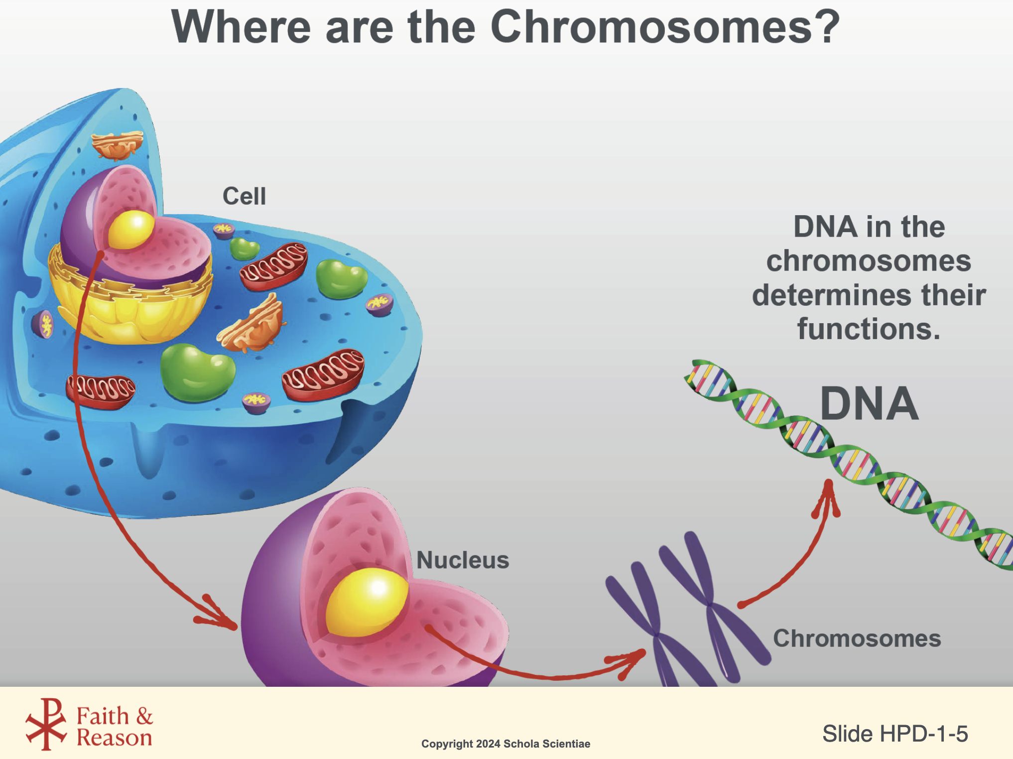
- Chromosomes are made of DNA, which carries the instructions for how every cell functions, divides, and develops.
Background Reading (Readings open in a new window):
Relevant Concept Slides (Click to enlarge):
Why this matters:
Students will be modeling chromosome behavior using blocks or manipulatives. They must understand what chromosomes actually represent (bundles of DNA instructions) before they attempt to model changes in chromosome number.
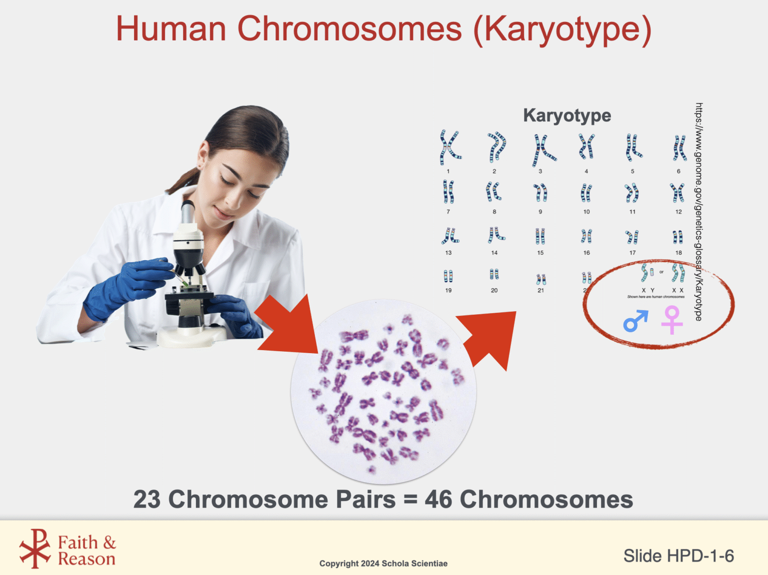
2. Humans Have 23 Chromosome Pairs — and This Matters for Growth
Key Idea:
Humans Have 23 Chromosome Pairs — and This Matters for Growth
Background Reading (Readings open in a new window):
Relevant Concept Slides (Click to enlarge):

Why this matters:
When students model mitosis, they must begin with the correct number of chromosomes. Miscounting the starting chromosome number is a common student mistake and leads to incorrect daughter cells later.
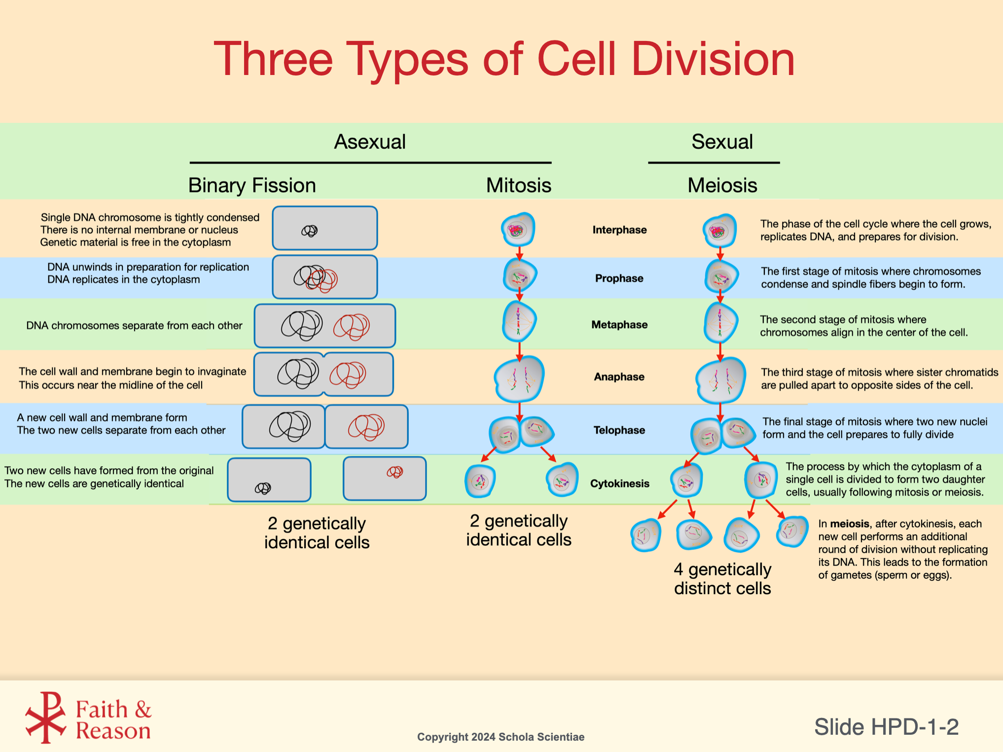
3. Mitosis Produces Genetically Identical Cells
Key Idea:
Mitosis ensures that each new cell receives a full set of DNA instructions—critical for growth, repair, and prenatal development.
Background Reading (Readings open in a new window):
Relevant Concept Slides (Click to enlarge):
Why this matters:
Students will model the four main stages (prophase → metaphase → anaphase → telophase).
Understanding the purpose of each stage helps them correctly manipulate their models and recognize errors in chromosome separation.
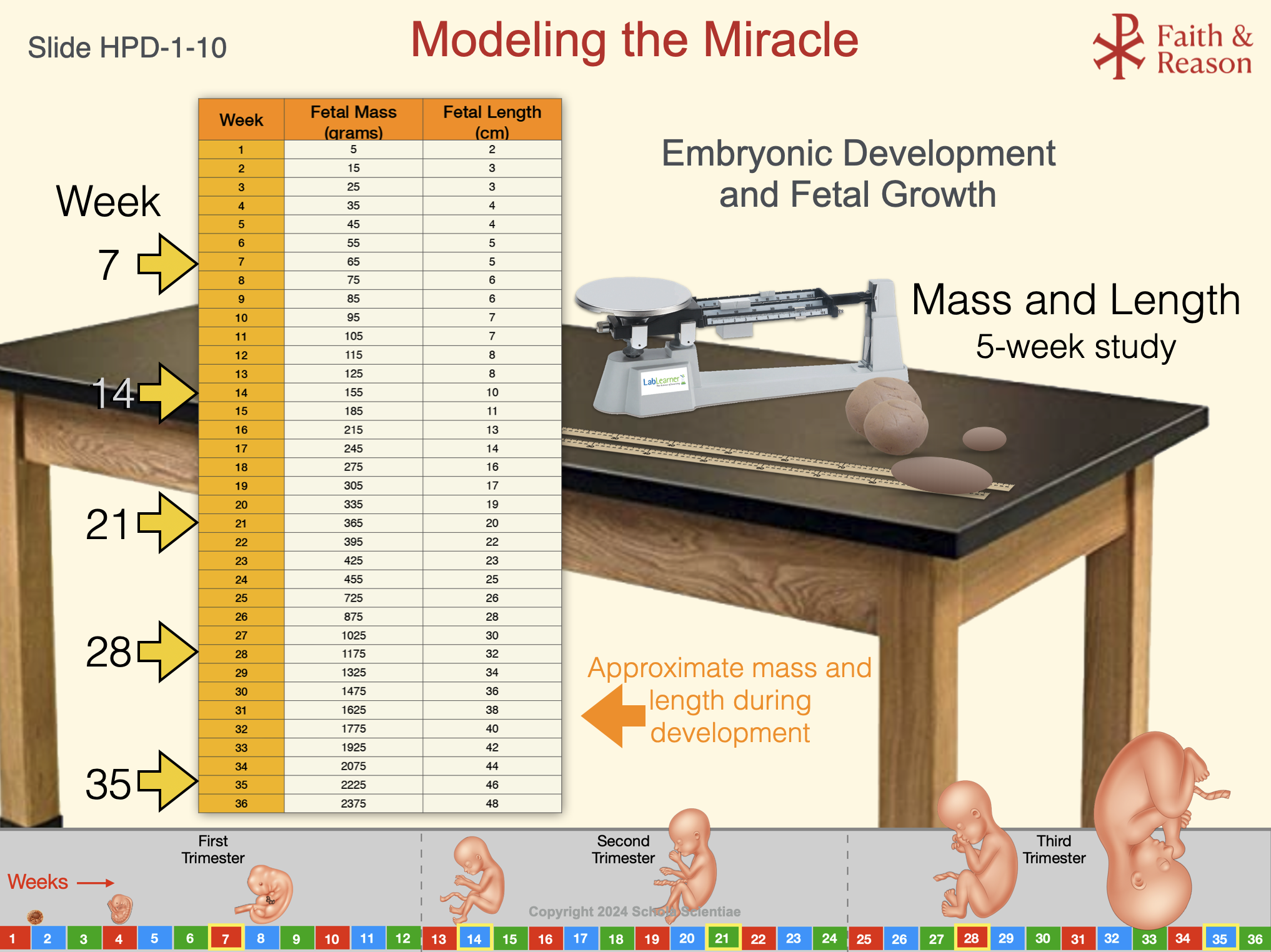
4. Mitosis Drives Massive Increases in Cell Number During Prenatal Development
Key Idea:
A human newborn has ~1.25 trillion cells. That number grows from one fertilized egg through countless rounds of mitosis.
Background Reading (Readings open in a new window):
Relevant Concept Slides (Click to enlarge):
Why this matters:
Students will connect the rate of mitosis to fetal mass and length using the Modeling the Miracle chart. Understanding that growth = cell division helps them interpret lab results and trimester modeling.
5. The Stages of Mitosis Must Occur Accurately
Key Idea:
If chromosomes do not separate correctly, the resulting cells may have too many or too few chromosomes.
Background Reading (Readings open in a new window):
Relevant Concept Slides (Click to enlarge):

Why this matters:
When students perform the Modeling the Miracle activity, they will model correct chromosome separation.
This reinforces why accuracy in mitosis is essential for healthy development.
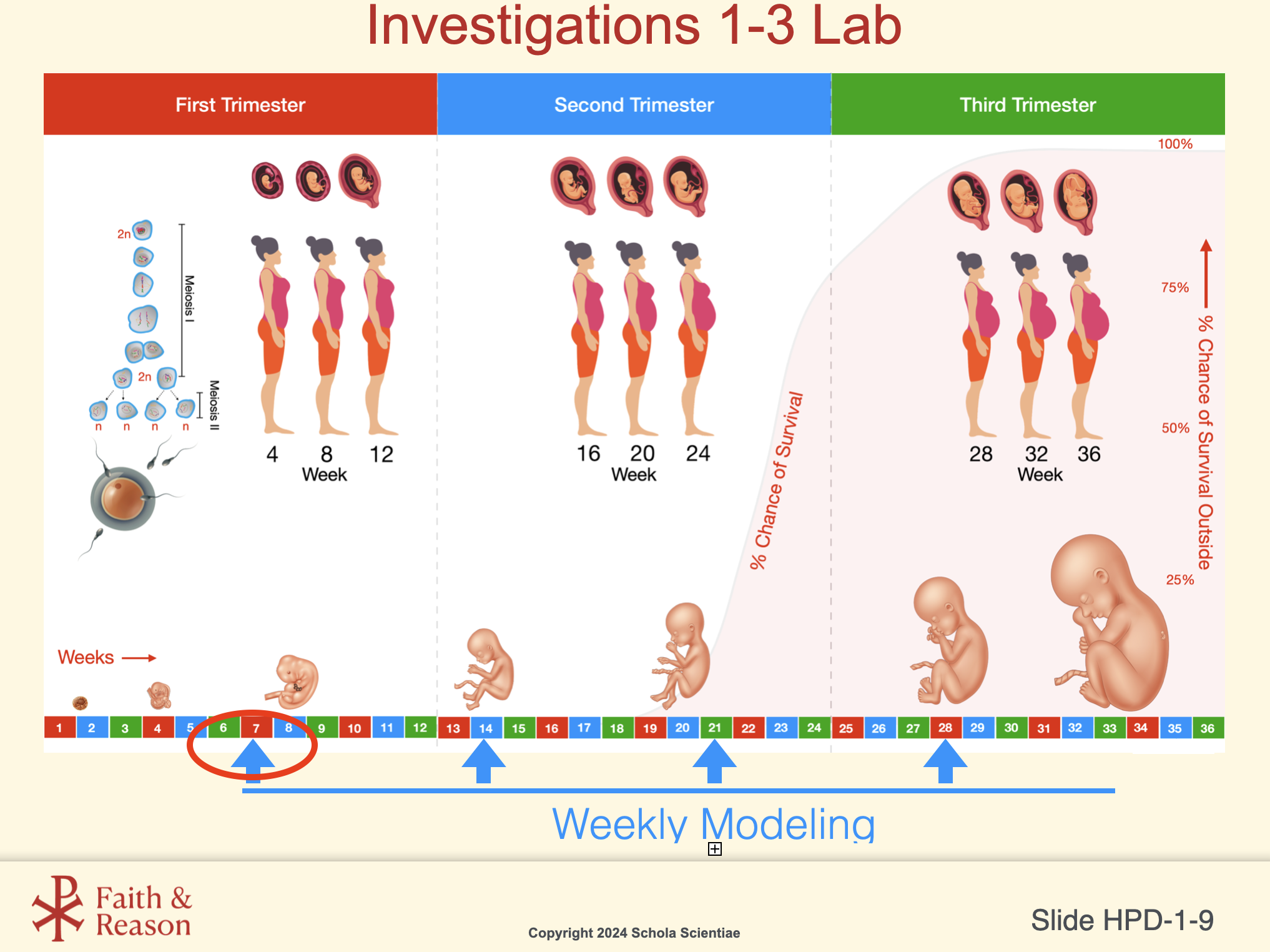
6. Growth Patterns Relate to Trimester Development
Key Idea:
Fetal growth follows predictable patterns. Understanding trimester-based changes helps students interpret the Modeling the Miracle data.
Relevant Concept Slides (Click to enlarge):
7. Students Will Apply All These Concepts in Lab
Key Idea:
The PreLab is not abstract — it is a roadmap for success in the Lab.
Students must be ready to:
- Begin with the correct chromosome number.
- Model chromosome duplication accurately.
- Model the stages of mitosis in correct order.
- Interpret fetal mass/length data as outcomes of mitotic cell increase.
Why this matters:
Students who walk into the Lab with these ideas clear will excel, make fewer construction mistakes, and produce more accurate interpretations.
Investigation Vocabulary:
Cell Division
Definition:
The process by which a cell reproduces by dividing into two new cells.
Teacher Notes — Why this matters:
This term anchors why students will model mitosis in HPD1.
Classroom Example:
Teacher asks: “Why does your body need cell division even right now?” Students connect it to repair and growth.
Chromosome (KROH-muh-sohm)
Definition:
A tightly coiled structure made of DNA that carries the genetic instructions for building and operating the body.
Teacher Notes — Why this matters:
Understanding chromosomes is essential for students to make sense of fertilization, cell division, genetic inheritance, and every stage of prenatal development.
Classroom Example — What this looks like:
“Every cell in your body has 46 chromosomes — the instruction sets that tell each cell what to do.”
Cytokinesis (SY-toh-kih-NEE-sis)
Definition:
The final stage of cell division in which the cytoplasm separates, forming two complete cells.
Teacher Notes — Why this matters:
Students will physically model this step using the Lab materials.
Classroom Example:
Teacher uses string to demonstrate the “pinching in” of animal cell cytokinesis.
DNA
Definition:
The molecule inside chromosomes that contains all the genetic information needed for growth, repair, and development.
Teacher Notes — Why this matters:
Students must understand DNA before they can understand how chromosomes replicate and why accurate copying is critical during mitosis.
Classroom Example — What this looks like:
“This long molecule has the instructions that make you you.”
Embryo (EM-bree-oh)
Definition:
An early stage of human development before major organs are formed.
Teacher Notes — Why this matters:
This lays groundwork for understanding early prenatal growth and the weekly modeling activity.
Classroom Example:
Teacher asks students to predict: “At which week would the embryo begin looking more like a fetus?”
Fetus (FEE-tus)
Definition:
The developing human from about week 9 until birth, when major organs have begun forming.
Teacher Notes — Why this matters:
Students must distinguish embryo vs fetus to interpret the weekly fetal mass and length table.
Classroom Example:
Teacher displays Week 14 vs Week 21 models and asks: “What growth patterns do you notice?”
Karyotype (CARE-ee-oh-type)
Definition:
An organized image of all the chromosomes in a cell, arranged in pairs.
Teacher Notes — Why this matters:
Understanding the normal set of 23 pairs sets up later lessons on meiosis and inheritance.
Classroom Example:
Teacher shows two karyotypes and asks: “Which one is male and how do you know?”
Mitosis (my-TOE-sis)
Definition:
The process in which a single cell divides to form two genetically identical cells.
Teacher Notes — Why this matters:
Growth, tissue repair, and cell replacement all depend on mitosis. Students must understand how chromosomes copy and separate accurately.
Classroom Example:
During a review, teacher asks: “Show me—what happens in metaphase?” Students point to chromosomes lining up in the center.
Nucleus (NOO-klee-us)
Definition:
The membrane-bound structure in the cell that houses the chromosomes.
Teacher Notes — Why this matters:
Students often forget that mitosis is division of the nucleus, not the entire cell.
Classroom Example:
Teacher taps a diagram: “Where is mitosis actually happening?” Students point to the nucleus.