
Teacher Portal
Investigation 4: Concepts
Navigate:
Once the slide presentation is launched
- use your left and right arrows to advance or go back in the slide presentation, and
- hover your mouse over the left edge of the presentation to get a view of the thumbnails for all the slides so that you can quickly move anywhere in the presentation.
- Click HERE to launch the slide presentation for the CELL.
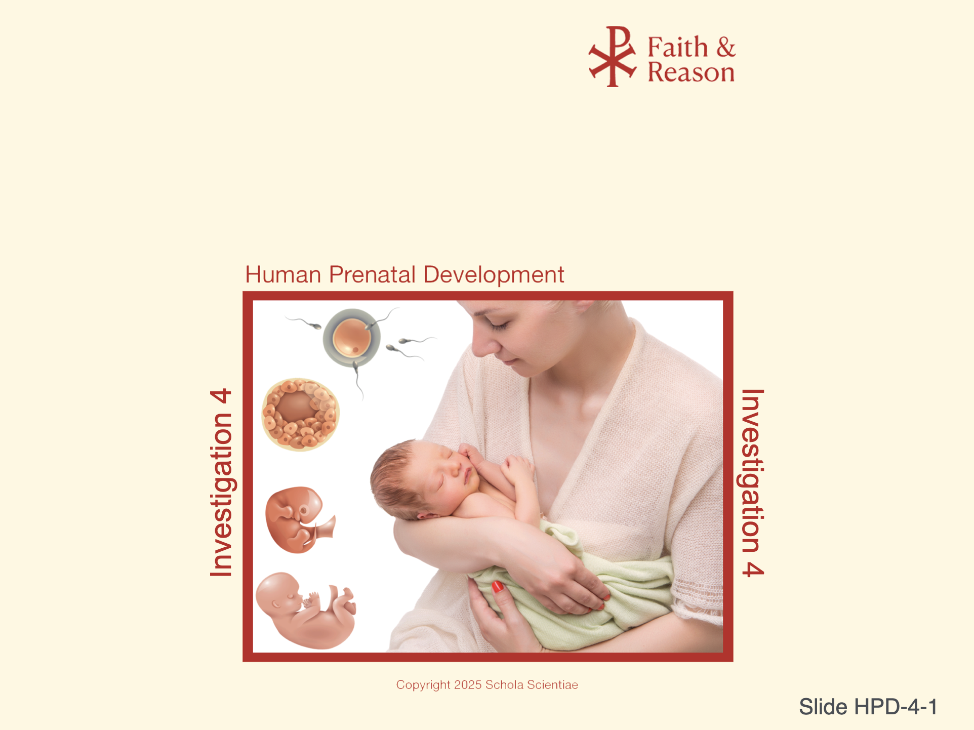
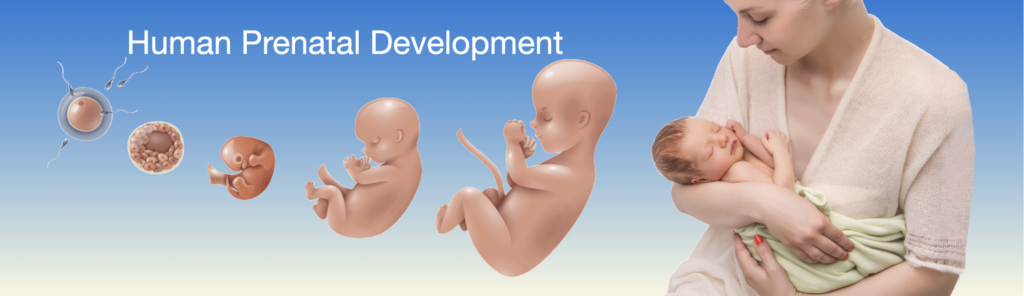
SLIDE HPD-4-1
This slide introduces Investigation 4 of the Human Prenatal Development CELL. By this point in the series, students have studied the earliest stages of human development—from fertilization and cell division through embryonic growth and fetal changes. This final Investigation shifts focus to the later stages of development, including organ maturation, fetal viability, and birth.
The image brings the full journey into view: from fertilization to birth. By highlighting a newborn in the arms of its mother alongside key prenatal stages, the slide emphasizes the continuity of human life from a single cell to a fully formed infant.
This visual serves as a reflective moment for students to recognize how far development progresses over 9 months. It sets the tone for more detailed scientific and ethical discussions ahead, including fetal survival, organ system function, and anatomical comparison through dissection.
Discussion Questions and Answers
Q1: What does this slide show us about human development?
A1: It shows the full journey from fertilization through birth, helping us see that development is a continuous, organized process starting with a single cell.
Q2: Why might it be helpful to look back at the earlier stages now?
A2: Looking back reminds us of how complex and incredible development is—how much happens before birth and how each stage builds on the one before it.
Q3: How does this image help prepare us for what’s coming in this Investigation?
A3: It connects what we’ve learned so far with what we will study next—like how organs finish developing and how the body gets ready to survive outside the womb.
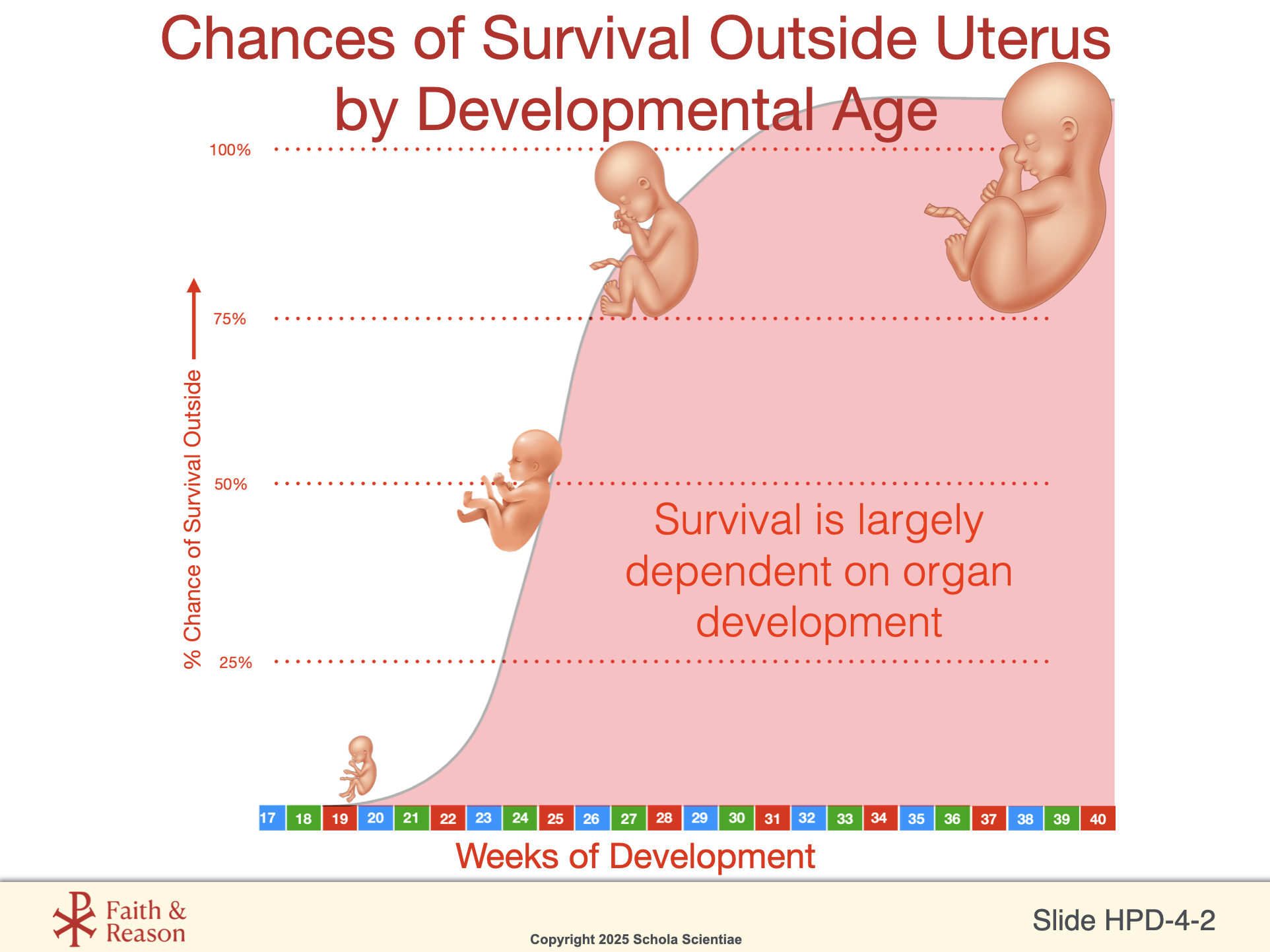
SLIDE HPD-4-2
This slide presents a graph showing the likelihood of a fetus surviving outside the uterus at different stages of prenatal development. It illustrates a steep increase in survival rates beginning around week 23 to 24, with near-total viability by week 36.
The key takeaway is that organ development—not just time—is what determines survivability. For example, even if a baby is born at 24 weeks, survival may still be difficult if the lungs or heart are not yet functional enough to support life outside the womb.
This is a critical moment to help students connect the concept of biological development with medical reality—and to consider how far modern medicine can go in supporting life outside the womb. It also introduces an ethical and practical lens to understanding why the later stages of pregnancy are so vital.
Discussion Questions and Answers
Q1: What does this graph show about fetal survival?
A1: It shows that the chance of survival increases rapidly after about 23 weeks, mostly because key organs like the lungs and heart are more developed.
Q2: Why can’t a fetus survive well before 20 weeks?
A2: Because many of its vital organs—especially the lungs—are not developed enough to function outside the uterus.
Q3: What is the main factor that allows a fetus to survive outside the womb?
A3: The development and function of vital organs, like the lungs, heart, and digestive system.
Q4: How do you think advances in medical care affect these survival rates?
A4: Medical advances like NICUs, ventilators, and better prenatal care have improved survival rates, especially for premature babies.
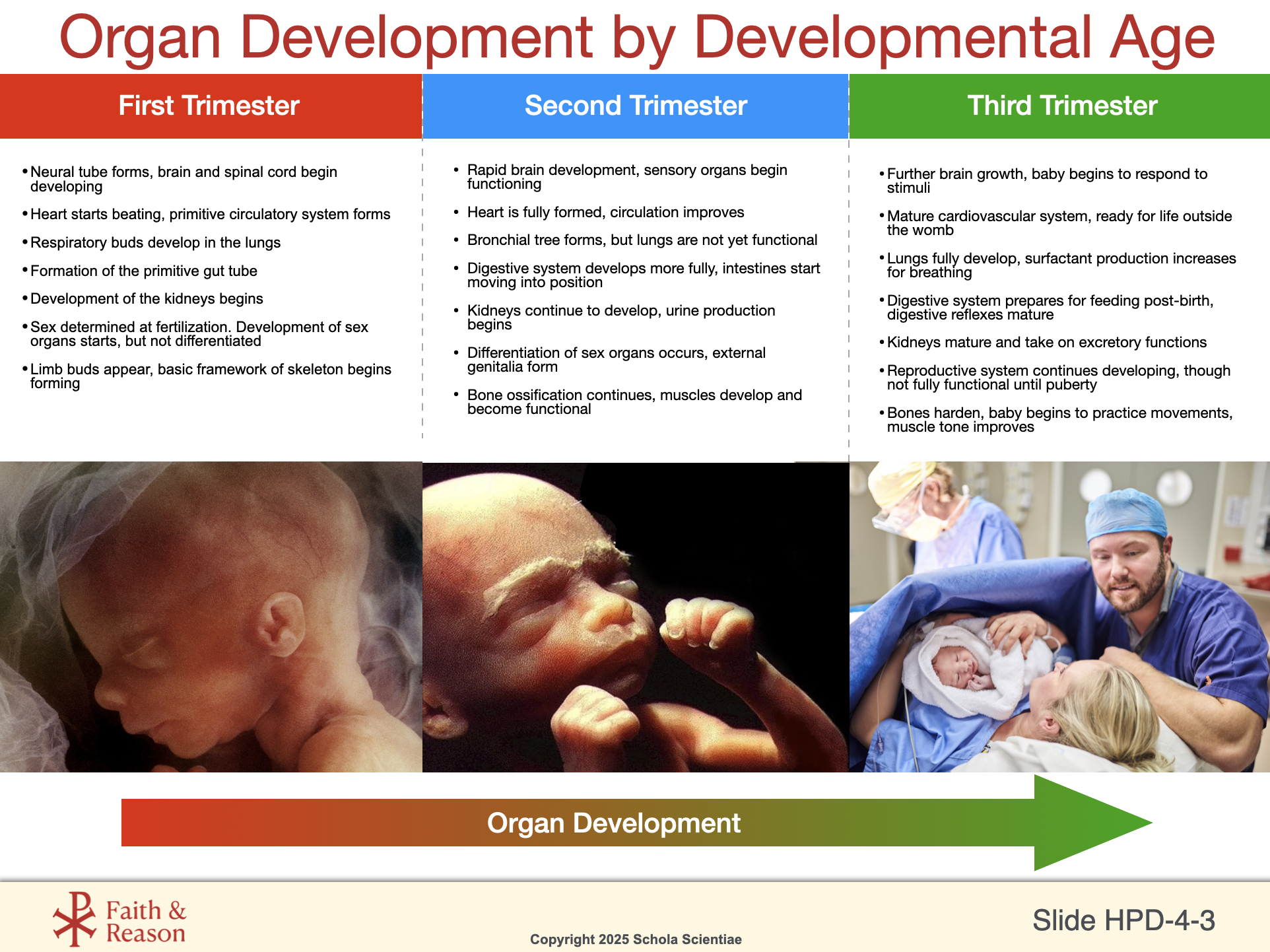
SLIDE HPD-4-3
This slide summarizes key organ development milestones during each trimester of pregnancy, helping students visualize how different body systems form over time. From the early creation of the neural tube and heartbeat in the first trimester, to functional kidneys and a growing brain in the second trimester, and finally to lungs preparing for breathing and bones strengthening in the third trimester, each stage builds on the one before.
This slide offers a chance to connect growth to survival—many of the organs listed here explain why survival increases as pregnancy progresses. For example, a fetus cannot survive if the lungs are not ready to breathe air, or if the heart and kidneys cannot regulate internal systems.
The images along the bottom help reinforce the connection between scientific stages and real fetal development. The final image of a newborn and the medical team reinforces the idea that birth is both the culmination of biological development and the beginning of independent life.
Discussion Questions and Answers
Q1: What are some important organs that begin developing in the first trimester?
A1: The brain, heart, lungs, digestive system, kidneys, and basic skeleton begin forming early in pregnancy.
Q2: What happens in the second trimester that helps the fetus survive better?
A2: Organs become more functional—like the kidneys starting to produce urine and the digestive system developing—plus the brain and muscles improve coordination.
Q3: Why is the third trimester so important?
A3: In the third trimester, organs like the lungs and digestive system mature enough to work outside the womb, and the baby gains fat, strength, and reflexes needed for birth.
Q4: Can you think of a system that begins developing early but isn’t fully functional until after birth?
A4: The reproductive system starts forming early but does not fully mature or become functional until puberty.
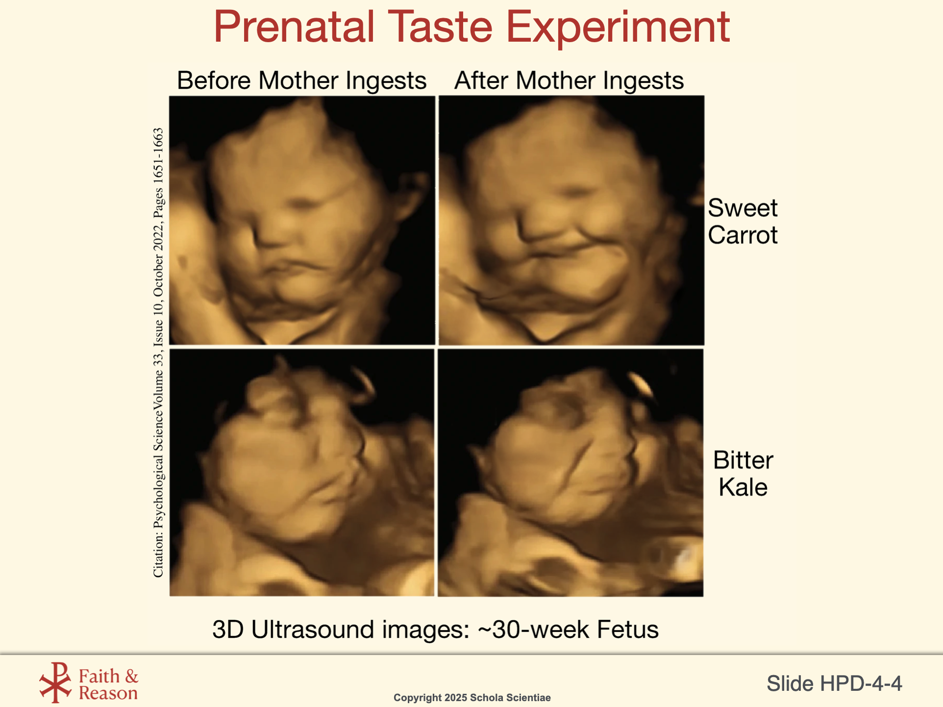
SLIDE HPD-4-4
Taste buds begin developing in the fetus and connecting to the brain around the 13th to 15th week of gestation, and by the third trimester, the fetus is capable of experiencing flavors.
How it works:
Amniotic fluid carries the flavors of the mother’s diet—like garlic, vanilla, carrots, or mint.
The fetus swallows this fluid regularly (up to a liter a day in late pregnancy).
Studies using ultrasound have shown that fetuses swallow more when the fluid tastes sweet (like after the mother ingests something sugary) and less when it’s bitter or unpleasant.
This particular slide highlights a fascinating study using 3D ultrasound imaging to show how fetuses react to different tastes. In the experiment, pregnant women were given either carrot juice (sweet) or kale juice (bitter). The resulting fetal facial expressions—smiles in response to carrots and frowns in response to kale—are not only striking but also deeply informative.
This tells us that by the third trimester, the sense of taste is already functional, and the fetus can detect flavors in the amniotic fluid, which changes based on what the mother eats. This provides an exciting and relatable way to connect sensory development with what you already know about flavor and food.
It also opens the door to deeper questions: Is this just a reflex? Or are the beginnings of preference and response taking root even before birth?
Discussion Questions and Answers
Q1: What did the fetus do when exposed to sweet carrot flavor?
A1: The fetus appeared to smile or show a relaxed facial expression.
Q2: How did the fetus react to bitter kale flavor?
A2: The fetus frowned or pulled back, showing a reaction of dislike.
Q3: How does the fetus detect taste?
A3: Flavors from the mother’s food pass into the amniotic fluid, which the fetus can taste by swallowing.
Q4: Why is this study important for our understanding of fetal development?
A4: It shows that senses like taste are active before birth, and that the fetus can respond to the outside world in subtle but meaningful ways.
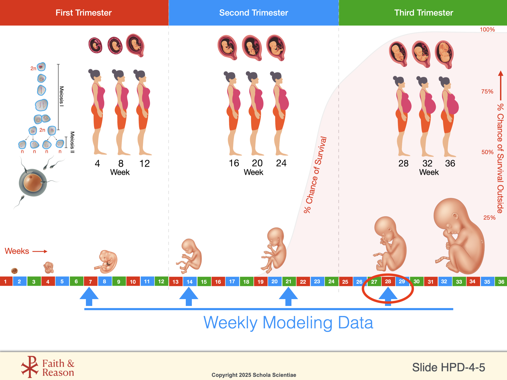
SLIDE HPD-4-5
This slide connects the students’ own Modeling the Miracle lab work to the broader timeline of prenatal development. It charts the growth of fetal mass over the course of pregnancy, showing the most dramatic acceleration during the third trimester—the final weeks before birth.
By comparing the mass at different weeks (e.g., 28 and 35 weeks), students can visualize how critical the last trimester is for survival, health, and strength at birth. It also helps them understand why premature babies often face challenges—they are born before this major growth surge has fully completed.
The table format encourages students to observe patterns in their own modeled data and relate it directly to biological growth, reinforcing scientific observation skills.
Discussion Questions and Answers
Q1: What trend do we see in fetal mass between weeks 28 and 35?
A1: There is a rapid and dramatic increase in mass, showing how important the final weeks are for growth.
Q2: Why is the third trimester sometimes called the “growth trimester”?
A2: Because the fetus gains most of its final mass and strengthens its organs in preparation for birth.
Q3: How does this modeling project help us understand real fetal development?
A3: By seeing the growth over time in our models, we better appreciate how quickly real fetuses grow and mature before birth.
Q4: Why do premature babies often need extra medical help?
A4: Because they are born before this major growth and organ strengthening is complete, so they need support until their systems can function independently.
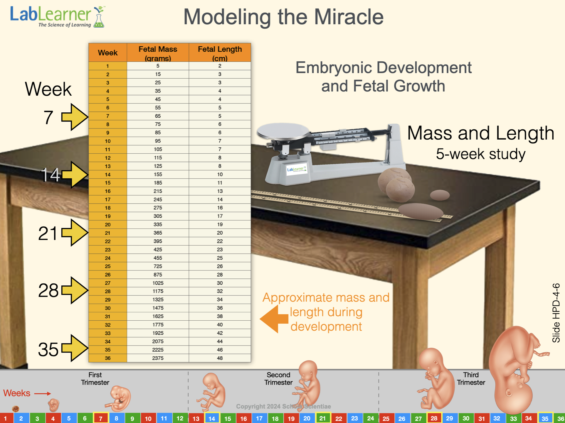
SLIDE HPD-4-6
This slide focuses on the hands-on Modeling the Miracle project and serves as a bridge between abstract scientific knowledge and physical experience. Students have been tracking fetal growth by increasing the mass and length of their clay models over several weeks. Now, they can see that their cumulative data mirrors real third-trimester growth rates.
This reinforces the importance of patient, careful observation over time—a fundamental principle of both science and medical practice. It also offers students a deeper, more tactile sense of the invisible biological changes occurring during fetal development.
This slide prepares students to transition from modeling to real biological structures in the upcoming dissection experience.
Discussion Questions and Answers
Q1: What did we use to model fetal growth in this project?
A1: We used clay to represent fetal mass and length, increasing it each week based on real developmental data.
Q2: Why is it important to measure growth week by week?
A2: It helps us see how growth is not constant but accelerates dramatically at certain points, especially in the third trimester.
Q3: How does modeling help us understand something we can’t easily see?
A3: Modeling gives us a physical way to experience and measure changes that are normally hidden inside the womb.
Q4: What does this project teach us about real scientific work?
A4: It shows that science often requires careful, consistent observation over long periods to understand important changes.
SLIDE HPD-4-7
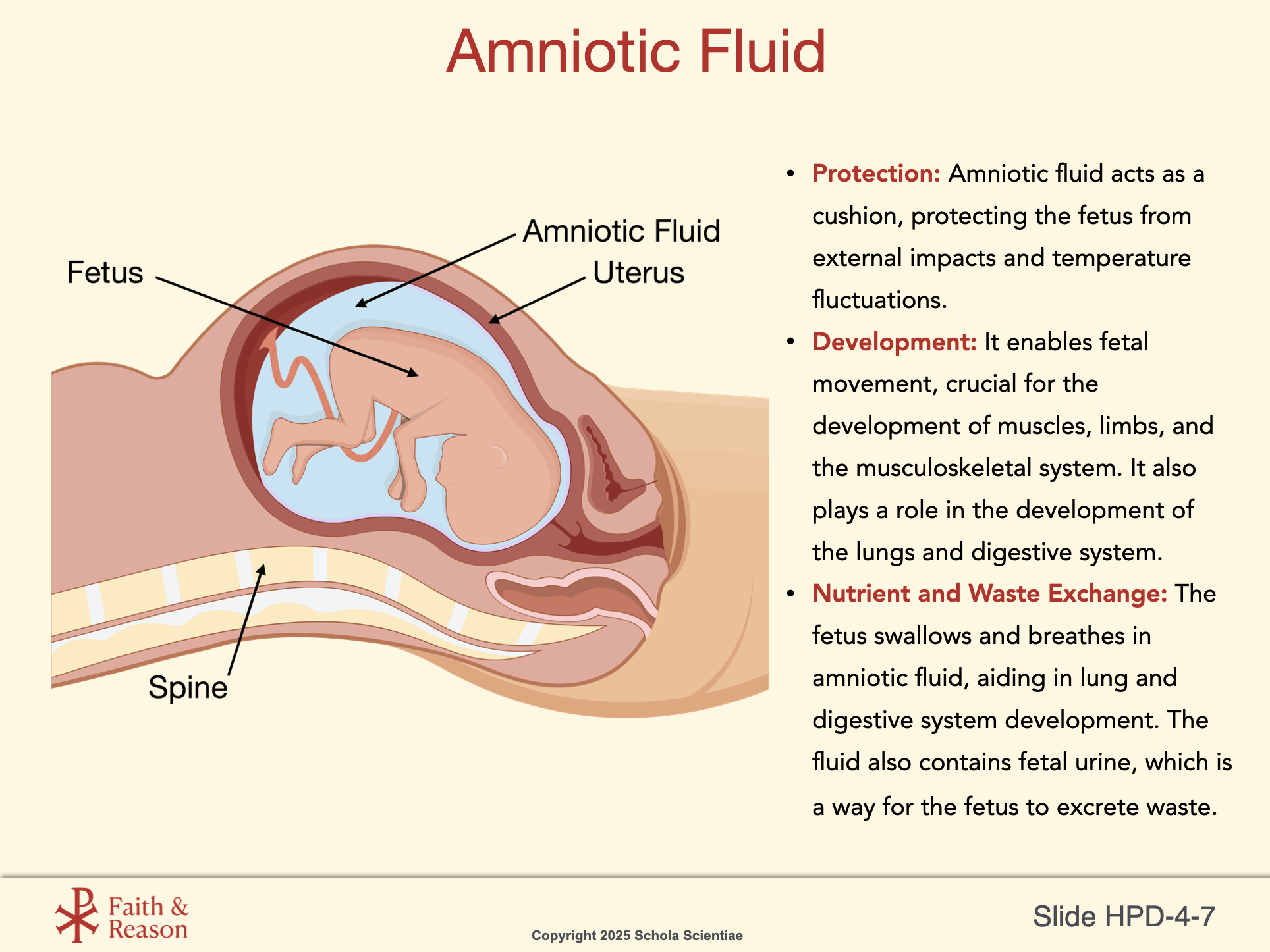
Amniotic fluid is the protective liquid surrounding a fetus during pregnancy. It cushions the baby, regulates temperature, and allows for movement, contributing to the healthy development of the fetus’s lungs, digestive system, and musculoskeletal system. It’s also a key indicator of fetal well-being, with its volume and composition monitored during prenatal care.
Key Functions of Amniotic Fluid:
- Protection: Amniotic fluid acts as a cushion, protecting the fetus from external impacts and temperature fluctuations.
- Development: It enables fetal movement, crucial for the development of muscles, limbs, and the musculoskeletal system. It also plays a role in the development of the lungs and digestive system.
- Nutrient and Waste Exchange: The fetus swallows and breathes in amniotic fluid, aiding in lung and digestive system development. The fluid also contains fetal urine, which is a way for the fetus to excrete waste.
The video below shows an ultrasound of a fetus. Notice its movements! This type of movement helps develop and strengthen its muscles in preparation for birth. You can also clearly see the beating fetal heart in this ultrasound.

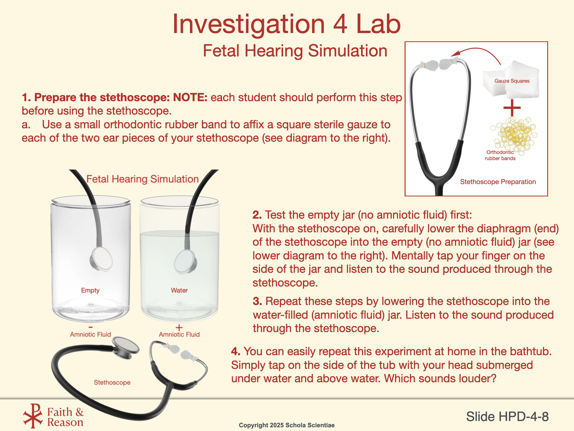
SLIDE HPD-4-8
Fetal Hearing Simulation: Sound Transmission in Amniotic Fluid
This investigation models how a fetus begins to hear during the third trimester while surrounded by amniotic fluid. By using a stethoscope and comparing sound transmission through air (an empty jar) versus water (a water-filled jar), students discover that sound travels more efficiently through fluid. This mirrors the fetal environment, where sound vibrations—especially from the mother’s voice—are transmitted through the amniotic fluid to the developing ears. This hands-on activity reinforces key physics concepts of sound conduction while connecting directly to human prenatal development.
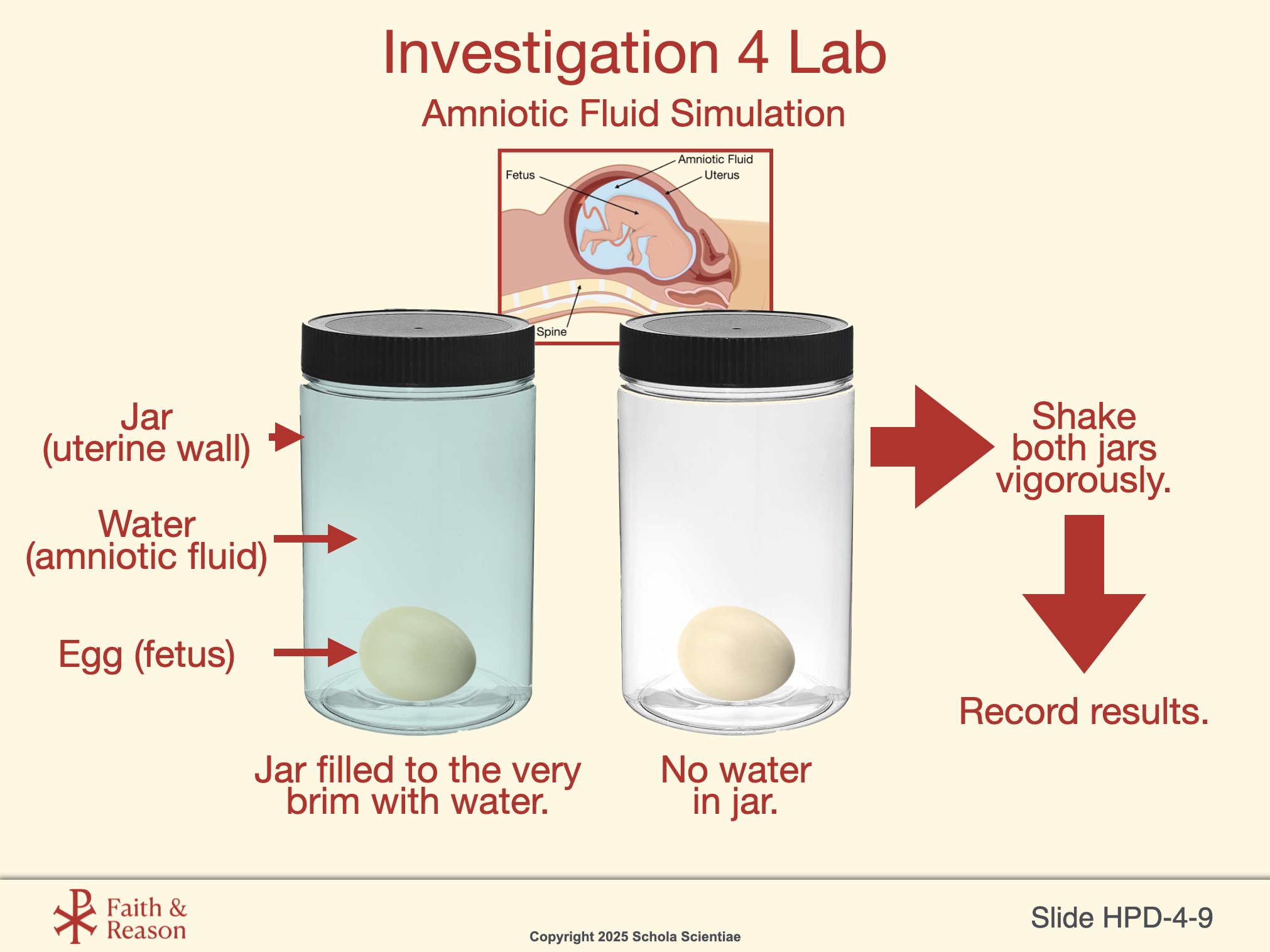
SLIDE HPD-4-9
This demonstration illustrates how amniotic fluid protects the fetus inside the womb. Students compare how physical impact affects an object (the egg) in two environments: surrounded by fluid versus air.
Setup: Two sealed jars each contain a raw egg. One jar is completely filled to the very brim with water (representing amniotic fluid), while the other is left dry in an empty jar. After sealing both jars tightly, students shake them vigorously and observe the outcome.
Scientific Principle: The jar filled with water disperses the kinetic energy of shaking, protecting the egg from damage. The dry jar transmits more force directly to the egg. Amniotic fluid serves as a cushion, absorbing and distributing mechanical forces to protect the developing fetus.
Visual Connection: The anatomical diagram helps students visualize the protective environment inside the uterus and how closely this experiment mirrors that structure.
Discussion Questions and Answers:
Q1: What do you think the water inside the womb (amniotic fluid) does for the developing baby?
A1: It likely helps cushion the baby and keep it safe from bumps or movements. It might also help the baby float and move more easily.
Q2: What advantage is there for a fetus to develop in fluid instead of just in air?
A2: Fluid helps support and protect the baby as it grows. It also helps the baby stay at a steady temperature and might allow it to move and develop its muscles more naturally.
Q3: If there were no fluid in the womb, what problems might that cause?
A3: The baby might get hurt more easily from any bumps or pressure. It could also have trouble moving or developing certain parts of its body properly.
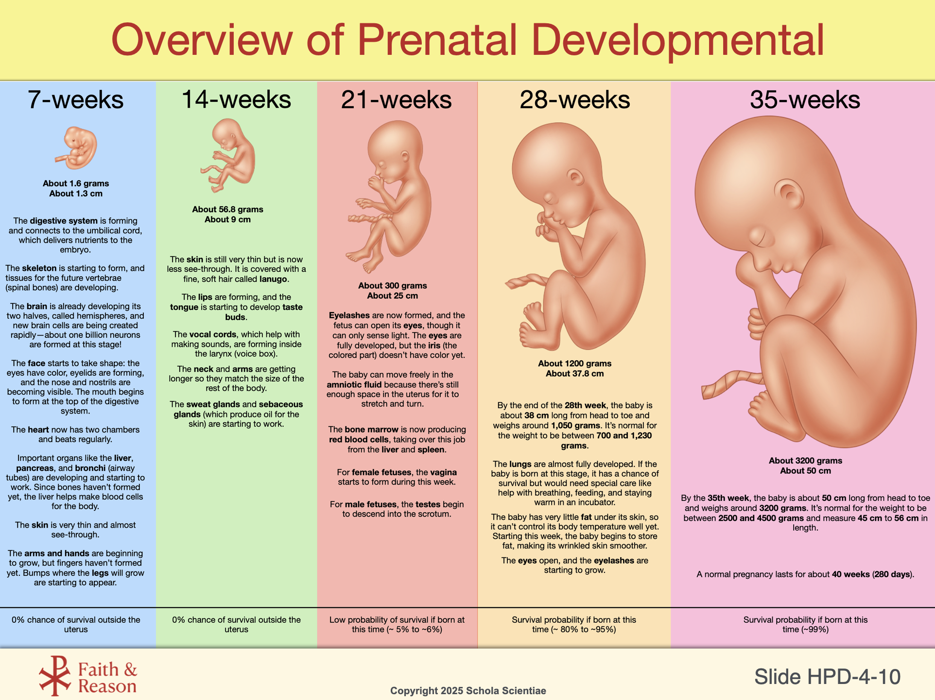
SLIDE HPD-4-10
This slide brings the entire Human Prenatal Development CELL to a close by presenting a timeline of development from weeks 7 to 35. It summarizes the major physical and functional changes students have explored in Investigations 1–4, including early cell division, organ formation, sensory development, and preparation for birth.
Students can now see how the data they’ve modeled, the organs they’ve learned about, and the scientific concepts they’ve discussed all come together in a real and measurable biological journey. Each week adds something essential: size, complexity, survival ability, and independence.
This is also an opportunity to invite reflection: how much have students learned—not just about human biology, but about patience, growth, and the hidden wonder of development?
This final scientific slide will also prepare students for a theological conclusion, where the fullness of this journey is celebrated in terms of human dignity and purpose.
Discussion Questions and Answers
Q1: What patterns do you notice across the weeks of development shown here?
A1: Growth speeds up over time, and major organs become functional as the fetus prepares for birth.
Q2: How does this timeline help explain why premature babies often need medical support?
A2: It shows that many systems (like the lungs) aren’t fully ready until the very end, so earlier birth can mean needing extra help to survive.
Q3: How did the Modeling the Miracle project connect to this timeline?
A3: Our weekly clay models matched these growth stages, helping us see how real and measurable fetal development is.
Q4: What is something new you now understand about human life before birth?
A4: Answers will vary, but students might say how complex it is, how much changes each week, or how early important features begin to form.