
Teacher Portal
Investigation 3: Concepts
Maternal-Fetal Interaction
Navigate:
Once the slide presentation is launched
- use your left and right arrows to advance or go back in the slide presentation, and
- hover your mouse over the left edge of the presentation to get a view of the thumbnails for all the slides so that you can quickly move anywhere in the presentation.
- Click HERE to launch the slide presentation for the CELL.
SLIDE HPD-3-1
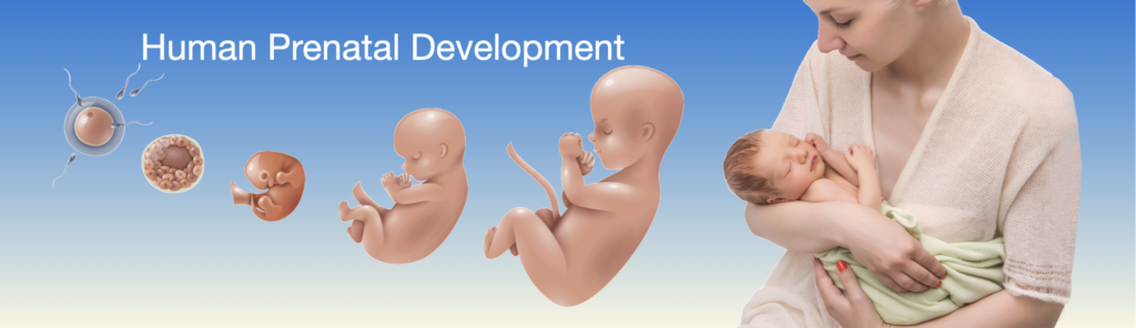
This title slide introduces Investigation 3 and sets a reflective tone. The developmental stages on the left—fertilization, early embryo, and fetus—provide a visual hook. Ask students what they already know about how a baby begins and grows before birth. This is a good moment to invite curiosity and connect the visuals to the personal and human side of biology.
Discussion Questions and Answers:
- Q1: What are the images on the left side of the slide showing?
- A1: They show the stages from fertilization to embryo to fetus.
- Q2: Why do you think the image of a mother holding a baby is included?
- A2: To help us understand that prenatal development leads to real people.
- Q3: What questions do you have about how a baby develops before birth?
- A3: (Open-ended; allow students to express wonder or curiosity.)
SLIDE HPD-3-2
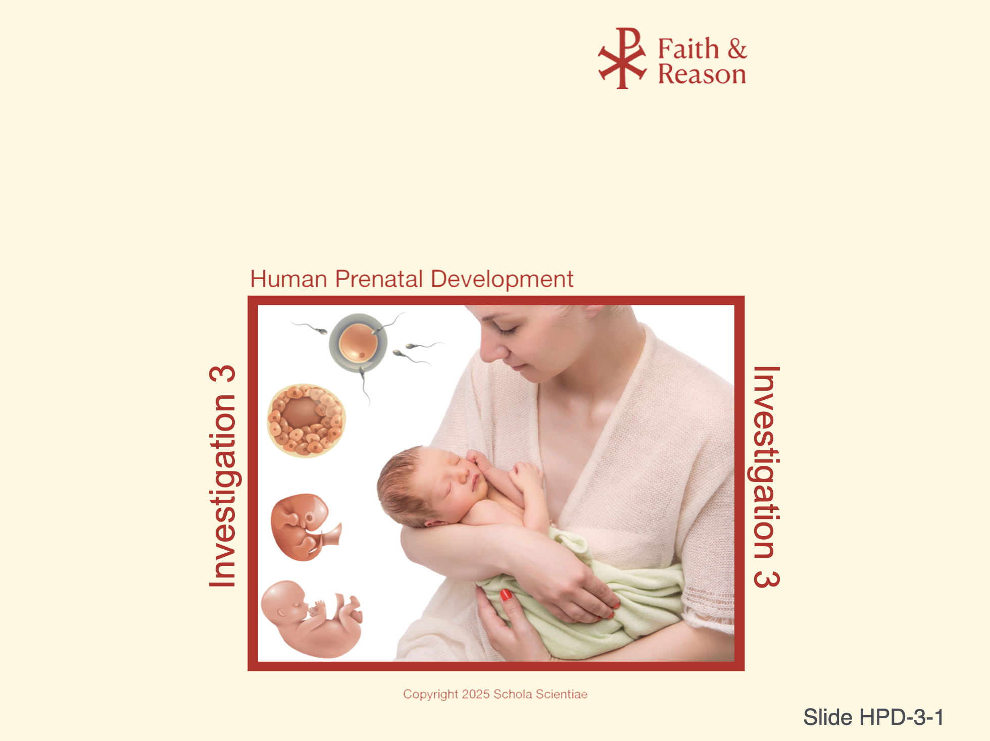
This is a review slide that lays the groundwork for understanding the biological process that begins human life. It distinguishes the roles of meiosis (producing haploid gametes) and mitosis (growth of the zygote). Emphasize that fertilization restores the diploid number and introduces genetic uniqueness.
Discussion Questions and Answers:
- Q1: What is the difference between haploid and diploid?
- A1: Haploid cells have half the DNA (n); diploid cells have a full set (2n).
- Q2: Why is fertilization necessary in creating a new human?
- A2: It combines DNA from both parents, starting a genetically unique human.
- Q3: What process allows the zygote to grow into more cells?
- A3: Mitosis is a type of cell division responsible for growth and tissue repair..
SLIDE HPD-3-3
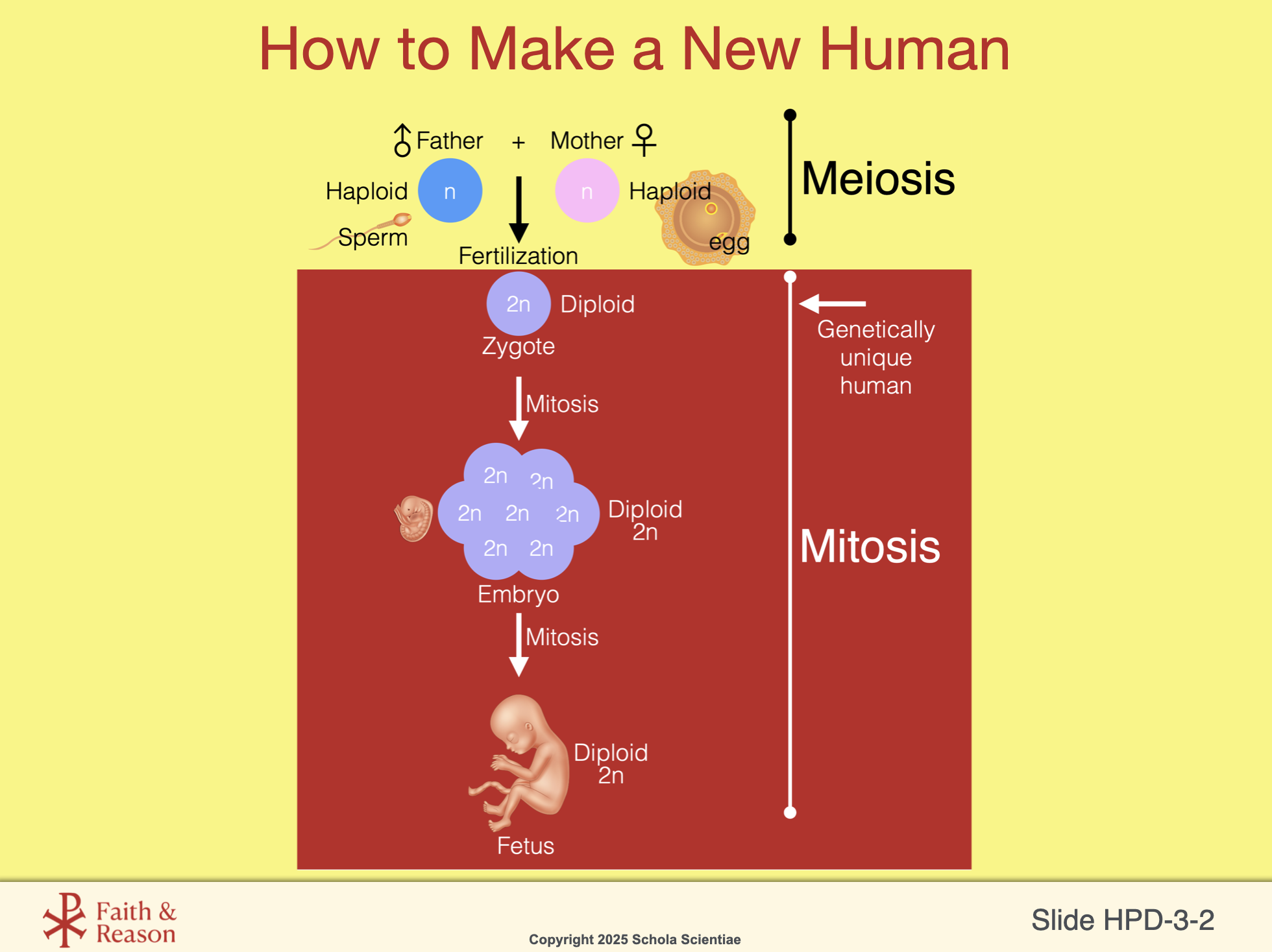
This is another review slide that zooms out to show the entire journey from gamete formation to birth. It’s essential to help students follow the timeline—from sperm and egg formation (meiosis) through fertilization, embryonic development (up to 9 weeks), fetal development, and ultimately birth. Reinforce how mitosis continues to drive growth throughout.
In addition, this review also emphasizes the early determination of the biological sex of the new embryo based on the father’s genetic contribution of either an X or a Y chromosome.
Discussion Questions and Answers:
- Q1: What is the difference between a zygote, embryo, and fetus?
- A1: A zygote is the single cell after fertilization, embryo is up to 9 weeks, fetus is after that.
- Q2: Where does mitosis take place in this timeline?
- A2: It happens after fertilization to grow the embryo into a fetus.
- Q3: What is the role of sperm and egg before fertilization?
- A3: They carry half the DNA and are made by meiosis.
SLIDE HPD-3-4
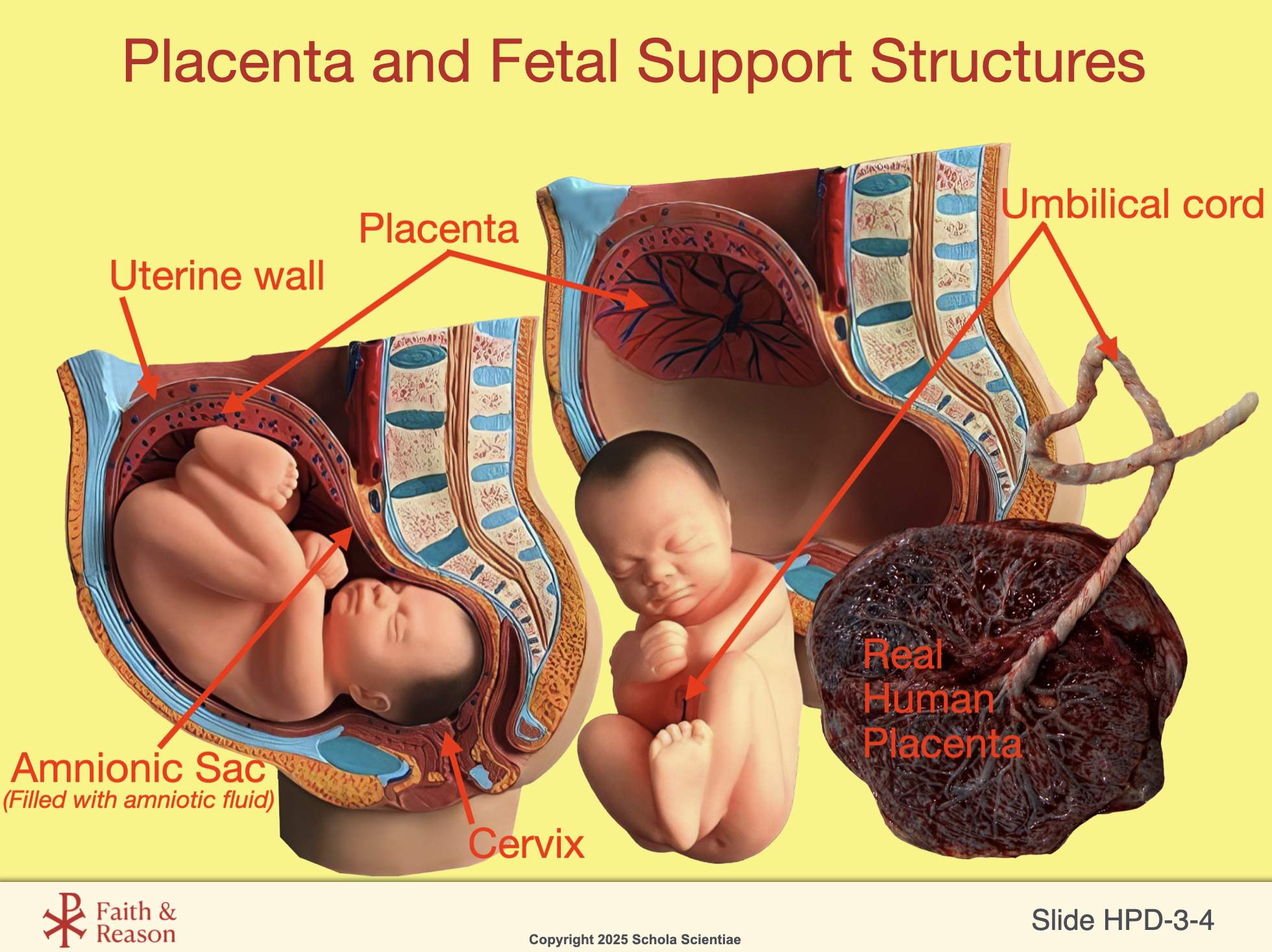
This anatomy-based slide helps students visualize how the fetus is supported inside the uterus. Explain the function of each structure: the amniotic sac for protection, placenta for nutrient and gas exchange, umbilical cord for transport, and cervix as the exit path.
Tell students that they can examine this identical model in lab (or have the model in class on Concept Day).
- Discussion Questions and Answers:
- Q1: What is the purpose of the amniotic sac?
- A1: To cushion and protect the fetus.
- Q2: How do nutrients and oxygen reach the fetus?
- A2: Through the placenta and umbilical cord.
- Q3: Is the baby floating inside the uterus?
- A3: Yes, in the amniotic fluid inside the sac.
SLIDE HPD-3-5
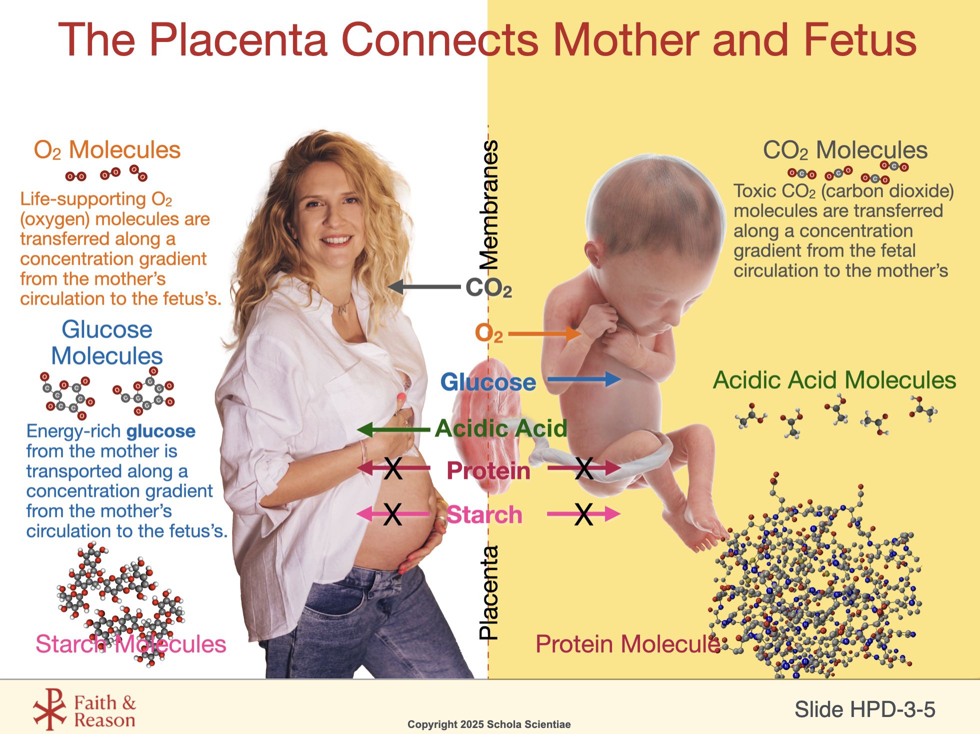
This slide shows how selective molecules pass through the placenta to support the baby’s growth. Point out that only small, specific molecules like oxygen and glucose can pass easily, while large molecules like starch and protein cannot. Toxic wastes like CO₂ and acidic waste flow from the baby back to the mother. This is a perfect setup for the filtration lab students will perform in the Investigation 3 lab.
Discussion Questions and Answers:
- Q1: What kinds of molecules can pass from the mother to the fetus?
- A1: Oxygen, glucose, and other small molecules.
- Q2: What happens to waste products like acidic acid and urea?
- A2: They pass from the fetus to the mother to be removed.
- Q3: Why can’t large molecules like starch go through?
- A3: They’re too big to pass through the placenta’s membranes.
SLIDE HPD-3-6
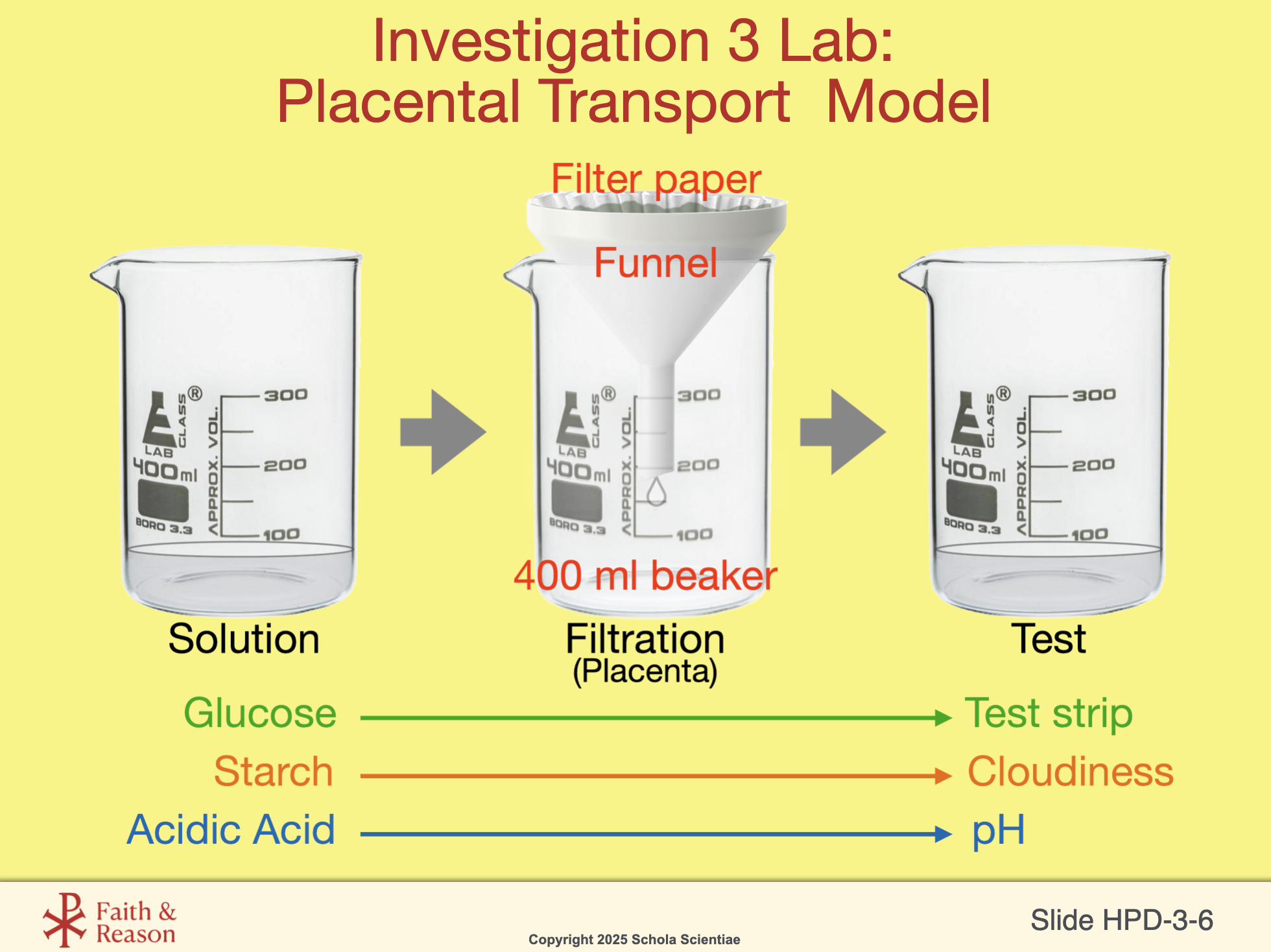
This slide previews the lab in which students will model the placenta using filter paper. Reinforce the idea that this is a “model”, not a perfect replica. Students will test which substances (glucose, acidic acid, starch) pass through and what that tells us about molecular size and diffusion in real biological systems.
Discussion Questions and Answers:
- Q1: What part of this setup represents the placenta?
- A1: The filter paper.
- Q2: What will we test for in the lab?
- A2: Whether glucose, starch, or acidic acid can pass through the filter (placenta).
- Q3: Why do we use different tests for different molecules?
- A3: Because each molecule needs a specific test to detect it.
SLIDE HPD-3-7
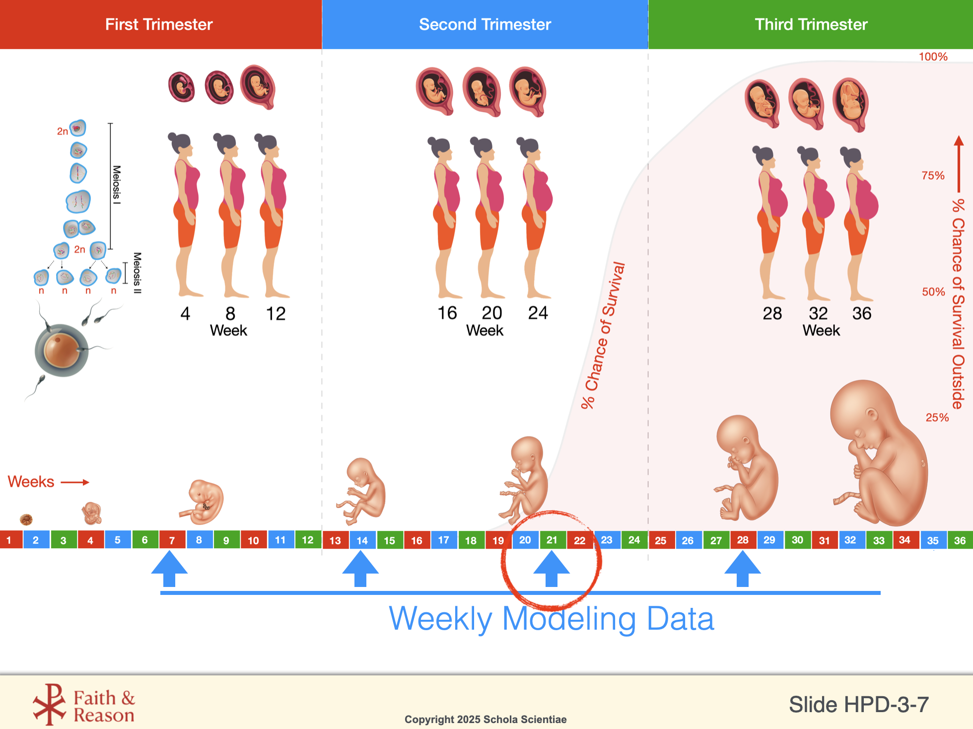
This developmental timeline helps students understand the week-by-week progression of the fetus and how survival chances improve. Use this to talk about preterm birth, fetal viability, and why the third trimester is so important. This slide also orients students for the weeks they’ll model.
Discussion Questions and Answers:
- Q1: What does the shaded red area mean on this chart?
- A1: It shows the chance of survival if the baby is born early.
- Q2: What do you notice about how the fetus changes between trimesters?
- A2: It grows in size and looks more like a newborn.
- Q3: Why is the third trimester important?
- A3: Because the baby is finishing development and preparing for birth.
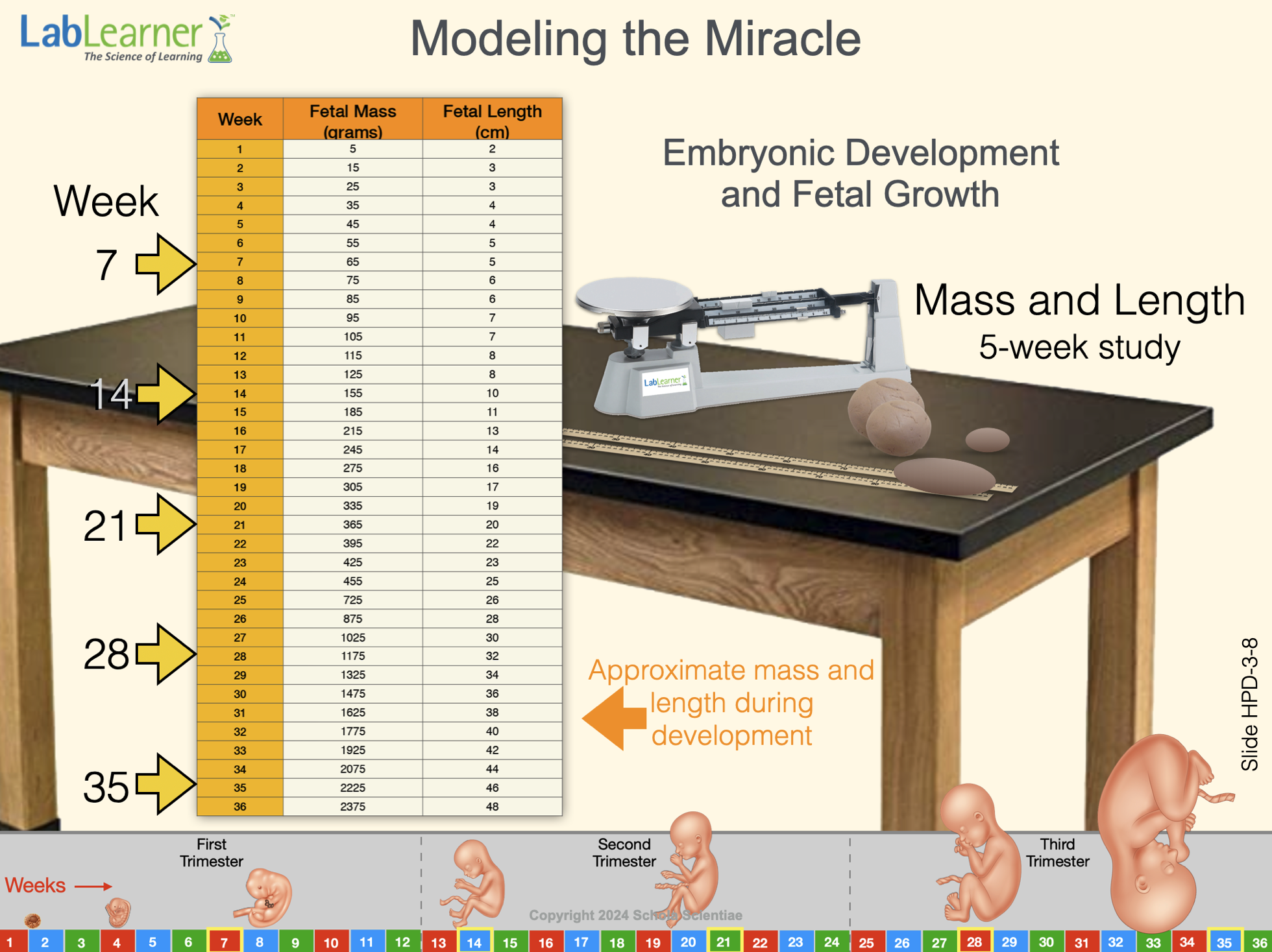
SLIDE HPD-3-8
This slide introduces the hands-on modeling activity where students track fetal growth in mass and length over time. In this Investigation, students will model the fetus at 21 weeks. Emphasize the precision of data and the awe-inspiring rate of growth during gestation.
Discussion Questions and Answers:
- Q1: What kind of data will we model in this lab?
- A1: The mass and length of a developing fetus.
- Q2: Why does the fetus grow more rapidly in the later weeks?
- A2: Because its organs are developing, and it’s getting ready to live outside.
- Q3: What tool will we use to track this growth?
- A3: A scale and a ruler to measure clay models.